Imaging tests are non-invasive procedures that provide pictures of the internal structures of your body. Common imaging tests include:
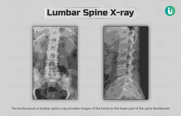
X-rays: In this test, the patient is asked to stand, sit or lie in front of a machine that emits x-rays. X-rays are invisible and intangible radiations which can pass through the body. However, as they travel through the human body, some amount of x-rays are absorbed by dense body tissues, like bones whereas soft tissues like organs do not absorb any. Whatever remains is then captured by a detector placed on the other side of the patient’s body, which then helps produce images of the part of the body being tested. So while bones would appear dark grey in an X-ray film, lungs appear black.
X-rays can be used to see both bones and soft tissues. An x-ray may be done to look for tooth and bone problems or injuries, lung problems, breast cancer (x-ray mammogram) and to check for the presence of metal objects stuck in the body like ingested coins. X-rays are also used to guide surgical procedures like to check for the proper placement of metal implants in joints or bones and in coronary angioplasty to guide the balloon into the right blood vessel.
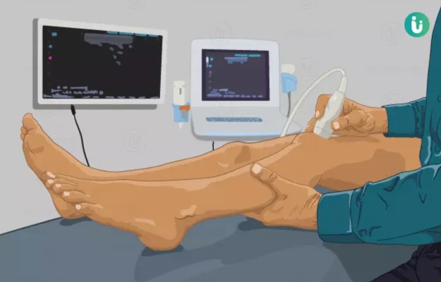
Ultrasounds: An ultrasound test uses a tiny probe to send sound waves into the concerned area of the patient’s body. These waves hit body tissues and bounce back to the probe, which then transfers the information to a computer to make an image of the tissue being tested. The images can be seen in real-time and can also be printed for later evaluation. Ultrasound tests are much safer than x-rays since they don’t involve any radiation exposure. Also, these tests can help visualise real-time changes in the body such as the movement of the fetus in the womb and blood flow through an artery (Doppler ultrasound).
Depending on the condition that needs to be checked, the probe is either moved over the patient’s body (external ultrasound) or inserted into their body (internal ultrasound). An endoscopy ultrasound is one where the ultrasound probe is attached on the tip of a long rod (an endoscope) and is passed into the patient’s body to take images.
Ultrasound can be used to check for problems with the structure or function of an organ. Doctors usually order an ultrasound for conditions like fibroids, stones, cysts, tumours and lumps in the body. High intensity focussed ultrasounds are a type of therapeutic ultrasounds in which very high-intensity sound waves are used to generate heat and clear blood clots, treat uterine fibroids or tumours in the body.
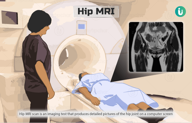
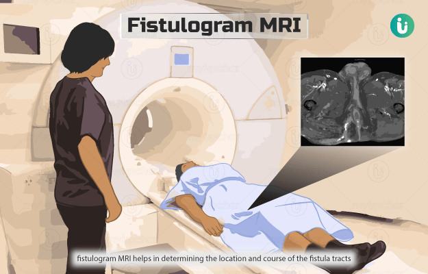
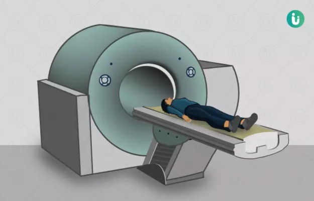
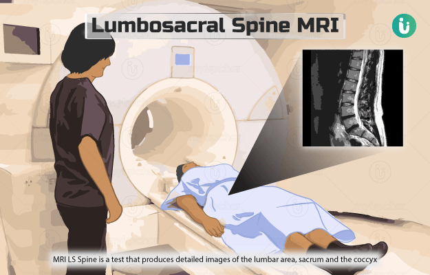
MRI: Magnetic resonance imaging or an MRI scan uses a combination of a magnetic field and radio waves to generate pictures of the internal structures of the body. An MRI machine is like a tunnel, the patient lies on the MRI table which then slides into the tunnel. During the scan, the machine usually makes a lot of noise and the patient is given headphones to mute the sound. The technician stays in an adjoining room but the patient can contact him/her through an intercom.
For claustrophobic patients, some places also offer a reclined open and a standing open MRI, where the patient either sits or stands in the MRI machine. MRI scans are used to check any body part including bones, joints, and soft tissues. This imaging test can also help check the brain function in real-time.
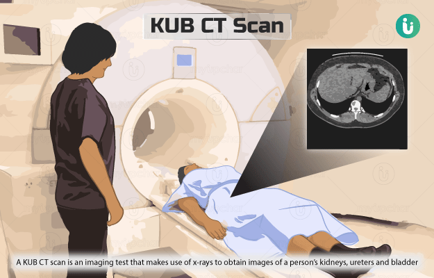
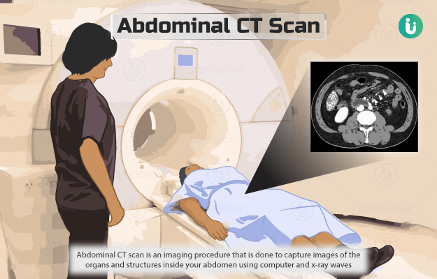
CT scan: A Computed Tomography (CT) scan also uses x-ray radiations to produce images of internal structures of the body. However, a CT scan machine has a circular scanner that rotates around the patient’s body while emitting and collecting x-rays and creating tomographic images (tiny slice images) of the patient’s body. A computer attached to the machine then collects all these slices to create a complete 3-D image.
Images obtained from a CT scan are hence much more detailed than a regular x-ray. Also, CT scans can be done to visualise any part of the body including soft tissues. Some of the things that a CT scan helps look for include injuries, tumours, blood flow, stroke and conditions like pneumonia and emphysema.
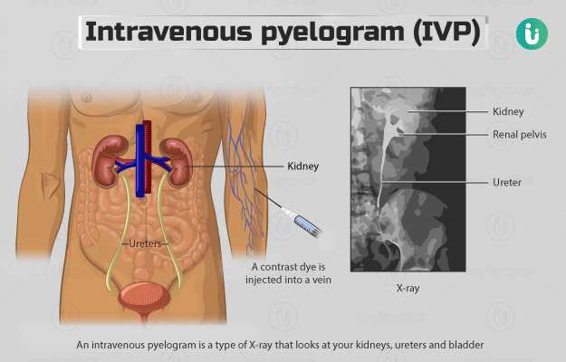
Contrast vs non-contrast imaging tests: A contrast dye is sometimes injected or given orally to patients before undergoing an imaging test. These dyes are taken up by tissues that need to be examined, which makes it appear a bit different than the surrounding tissues and is hence more clearly visible.
After the test, the dye slowly gets flushed out of the body.
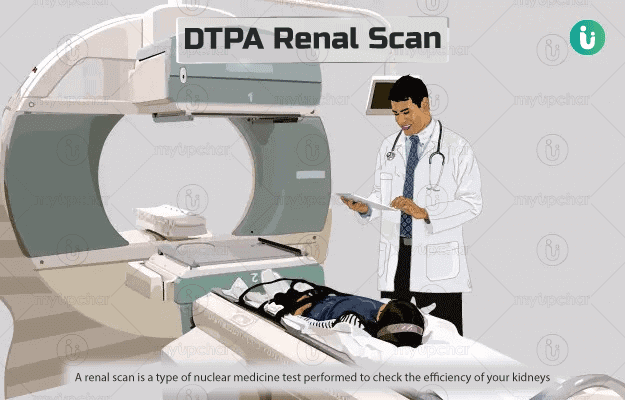
PET scan: A positron emission tomography or PET scan involves the administration of a radioactive tracer to the patient either through injection, inhalation or oral route. This tracer is absorbed by the tissues, leading them to emit gamma rays inside your body. A PET scan machine, which looks a lot like a CT scan machine, picks up the radiations emitted by these tissues and present a complete image of the concerned tissue on the computer screen. A PET scan is much more sensitive than an MRI and CT scan as it can identify problems while they are still at the cellular level. PET scan is most commonly done to check for cancer screening and diagnosis and to monitor the treatment of cancer. It is also used to diagnose conditions like Parkinson’s and epilepsy and coronary artery disease.


