What is a Computed Tomography (CT) scan of the whole abdomen? 

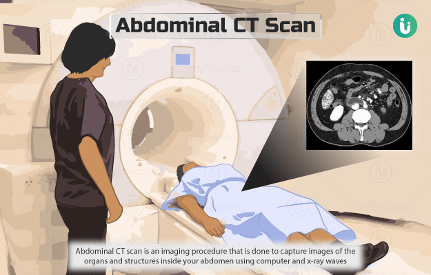
Computed tomography (CT) scan of the abdomen is an imaging procedure that is done to capture images of the organs and structures (blood vessels, muscles, and fat for example) inside your abdomen using computer and x-ray waves.

These images enable the healthcare provider to check for any abnormalities in your abdominal cavity.

During the scan, several x-ray beams move around your body (sometimes in a spiral path) as you pass through the CT scan machine and the radiation absorbed throughout the body is measured. A computer reads this data and produces two-dimensional images of the scanned area. 

CT scan provides detailed images of the internal organs and tissues. However, a contrast dye is sometimes administered into the body of the patient to help view the images more clearly. The dye binds to specific tissues and creates light and darker areas that makes it easier for the doctor or technician to differentiate them.

  1. Why is a CT Scan of the whole abdomen done?
  2. Who cannot have a CT Scan of the whole abdomen?
  3. How should I prepare for a CT Scan of the whole abdomen?
  4. How is a CT Scan of the whole abdomen done?
  5. How will a CT Scan of the whole abdomen feel like?
  6. What do the results of a CT Scan of the whole abdomen mean?
  7. What are the risks and benefits of a CT Scan of the whole abdomen?What are the risks and benefits of a CT Scan of the whole abdomen?
  8. What happens after a CT Scan of the whole abdomen?
  9. What are the other tests that can be done with a CT Scan of the whole abdomen?

A CT scan is performed to check for any abnormalities (such as tumour, infection or inflammation) related to the following organs in the abdominal area:

  • Liver
  • Spleen 
  • Kidney 
  • Bladder
  • Stomach 
  • Intestine 
  • Pancreas 
  • Adrenal gland (present on the top of kidneys)

Your healthcare provider may also order this test:

myUpchar doctors after many years of research have created myUpchar Ayurveda Urjas Capsule by using 100% original and pure herbs of Ayurveda. This ayurvedic medicine has been recommended by our doctors to lakhs of people for sex problems with good results.
Long Time Capsule
₹712  ₹799  10% OFF
BUY NOW

CT scans are usually safe. However, they should be avoided in case of:

You will be asked to wear loose, comfortable clothes and remove all the metal objects or jewellery before the test. The hospital staff may give you a gown to wear for the test. 

Fasting may be needed for a few hours before the test, especially if a contrast dye is used. Convey to your doctor if you are pregnant or allergic to contrast dye. 

Let your doctor know if you have asthma, diabetes, kidney diseases or thyroid or heart disorders. Diabetic people may be suggested taking their medicines at least two days before the test.

Your healthcare provider will ask you to sign a consent form. This form includes all the information about the risks associated with contrast dye.

The following procedure is performed for a CT scan:

  • You will be asked to lie down on the CT scanner table. During the test, this table will slide into the scanner. 
  • If a contrast dye is required, it will be injected into a vein in your arm or given to you in the form of a liquid to swallow before the test. 
  • The technologist will control the scanner from another room. He/she can communicate with you through the speakers inside the scanner. 
  • As the scanning procedure begins, the machine will pass x-ray waves through your body. 
  • The scanner will detect the x-rays absorbed by your body tissues and transfer them to an attached computer which will then interpret this data into images.
  • The scan takes anywhere between 15 minutes to half an hour. 
  • The technician will ask you to avoid movement during the scan as it may otherwise affect the final images. 
  • If an intravenous line has been placed for administering the contrast dye, it will be removed after the scan.

CT scan is a quick and painless imaging test. However, you may feel uncomfortable remaining still for the procedure. You may feel cold due to the air-conditioning temperature inside the room. When a contrast dye is injected, there may be a brief headache, flushing sensation, metallic taste in the mouth or vomiting. A mildly unpleasant taste may be felt when the contrast dye is swallowed.

An abnormal result can indicate the following conditions: 

 The benefits of the CT scan are as follows:

  • It is a non-invasive and painless procedure
  • It can detect bones, blood vessels and soft tissues simultaneously
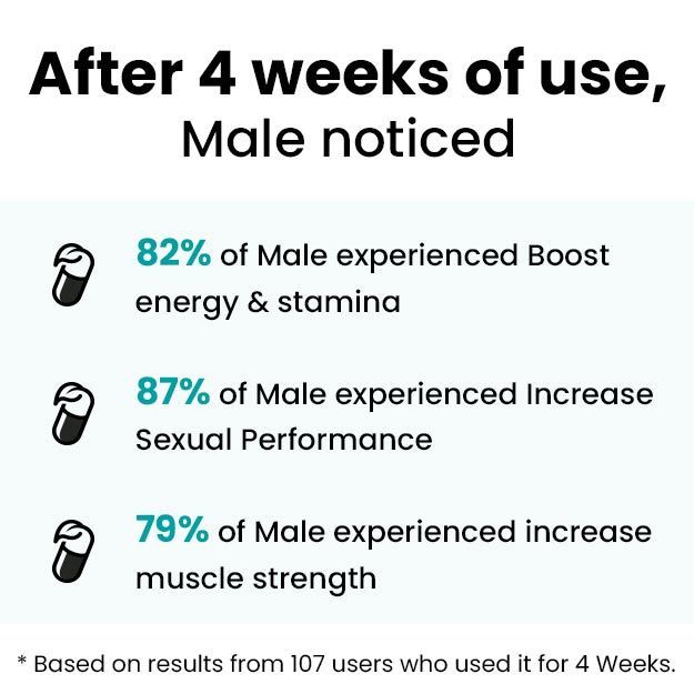
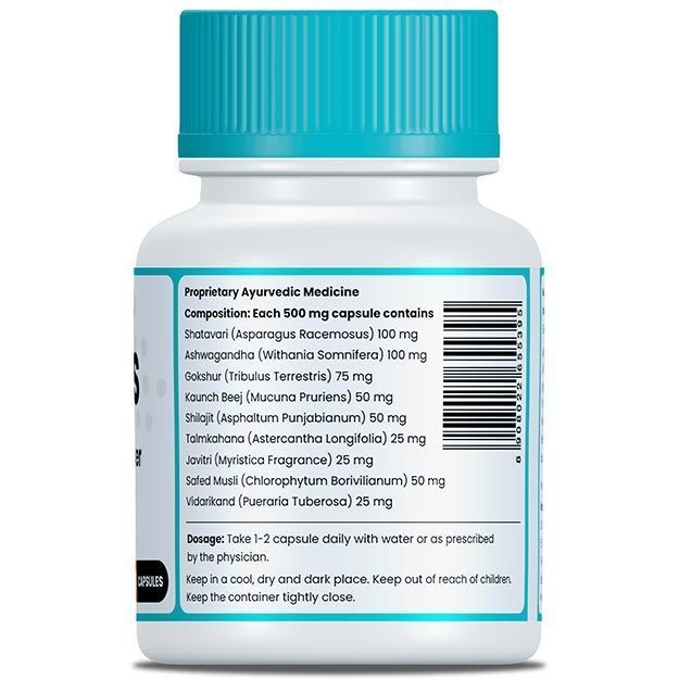
myUpchar doctors after many years of research have created myUpchar Ayurveda Urjas Energy & Power Capsule by using 100% original and pure herbs of Ayurveda. This Ayurvedic medicine has been recommended by our doctors to lakhs of people for problems like physical and sexual weakness and fatigue, with good results.
Power Capsule For Men
₹719  ₹799  10% OFF
BUY NOW

You can continue your regular diet and activities after the test. If a contrast dye is injected, your healthcare provider will check for any side effects such as itching, rash, swelling or difficulty in breathing. If you are consuming oral diabetic medicines, you may be asked to wait for 48 hours before resuming them.

Depending on the condition diagnosed, your doctor may order one of the following tests along with a CT scan of the whole abdomen:

Disclaimer: All results must be clinically correlated with the patient’s complaints to make a complete and accurate diagnosis. The above information is provided from a purely educational point of view and is in no way a substitute for medical advice by a qualified doctor.

References

  1. Johns Hopkins Medicine [Internet]. The Johns Hopkins University, The Johns Hopkins Hospital, and Johns Hopkins Health System; Computed Tomography (CT or CAT) Scan of the Abdomen
  2. Nemours Children’s Health System [Internet]. Jacksonville (FL): The Nemours Foundation; c2017; CAT Scan: Abdomen
  3. Jameson JL, De Groot LJ, de Kretser DM, et al, eds. Endocrinology: Adult and Pediatric. 6th ed. Philadelphia, PA: Elsevier Saunders; 2013
  4. Patel D, Nilubol N, Kebebew E. The management of pheochromocytoma. In: Cameron JL, Cameron AM, eds. Current Surgical Therapy. 12th ed. Philadelphia, PA: Elsevier Saunders; 2017:760-767
  5. National Cancer Institute. [Internet]. National Institute of Health. U.S. Department of Health & Human Services; Pheochromocytoma and Paraganglioma Treatment (PDQ®)–Health Professional Version.
  6. Smith KA. Abdominal pain. In: Walls RM, Hockberger RS, Gausche-Hill M, eds. Rosen's Emergency Medicine: Concepts and Clinical Practice. 9th ed. Philadelphia, PA: Elsevier; 2018:chap 24
  7. Wood CG, Berggruen S. Computed tomography of the solid abdominal organs. In: Gore RM, Levine MS, eds. Textbook of Gastrointestinal Radiology. 4th ed. Philadelphia, PA: Elsevier Saunders; 2015:chap 65
  8. Kim DH, Pickhardt PJ. Diagnostic imaging procedures in gastroenterology. In: Goldman L, Schafer AI, eds. Goldman-Cecil Medicine. 25th ed. Philadelphia, PA: Elsevier Saunders; 2016:chap 133
  9. Shaw AS, Prokop M. Computed tomography. In: Adam A, Dixon AK, Gillard JH, Schaefer-Prokop CM, eds. Grainger & Allison's Diagnostic Radiology. 6th ed. New York, NY: Elsevier Churchill Livingstone; 2015:chap 4
  10. Wang DQ-H, Afdhal NH. Gallstone disease. In: Feldman M, Friedman LS, Brandt LJ, eds. Sleisenger and Fordtran's Gastrointestinal and Liver Disease. 10th ed. Philadelphia, PA: Elsevier Saunders; 2016:chap 65
  11. Jackson PG, Evans SRT. Biliary system. In: Townsend CM Jr, Beauchamp RD, Evers BM, Mattox KL, eds. Sabiston Textbook of Surgery: The Biological Basis of Modern Surgical Practice. 20th ed. Philadelphia, PA: Elsevier; 2017:chap 54
  12. Barshak MB. Pancreatic infection. In: Bennett JE, Dolin R, Blaser MJ, eds. Mandell, Douglas, and Bennett's Principles and Practice of Infectious Diseases, Updated Edition. 8th ed. Philadelphia, PA: Elsevier Saunders; 2015:chap 78
  13. Lowe LH, et al. Unenhanced limited CT of the abdomen in the diagnosis of appendicitis in children: comparison with sonography. AJR Am J Roentgenol. 2001 Jan; 176(1): 31-5. PMID: 11133533.
  14. Diop AD, et al. CT imaging of peritoneal carcinomatosis and its mimics. Diagn Interv Imaging. 2014 Sep; 95(9): 861-72. PMID: 24631039.
Read on app
cross
Ask your health query from live doctors now!