A cannula is a tube that is inserted into the body to perform the function of delivering or removing any bodily fluid or taking samples. A cannula surrounds the inner or outer area of a needle (trocar) and increases the effective length of the needle by half the length of the original needle. It is commonly called an intravenous cannula (IV cannula). Peripheral venous cannulation (cannula inserted directly into the peripheral veins of the body) is the most common method to deliver intravenous therapy. The ultimate goal of intravenous management is to deliver treatment safely and effectively without any major discomfort or damage to tissues and without compromising venous access. This is especially important when long-term treatment is advocated. There are various indications and contraindications for peripheral cannulation; however, research suggests that intravenous cannulas are often inserted in patients when it can easily be avoided. In a recent study conducted in a sample of 1,000 patients admitted in general medicine wards, intravenous cannulas were absolutely idle in around 33% of the patients. An idle cannula is a cannula that is not used for 48 hours or more or without any prophylactic indication (when a cannula is inserted in anticipation of any requirement).

  1. Indications of IV cannulation
  2. Advantages of IV cannulation
  3. Contraindications of cannula insertion
  4. Types of cannulas
  5. Preparation before cannulation
  6. Materials required for IV cannulation
  7. Process of cannulation
  8. Complications of cannulation
Doctors for Cannulation

Following are some of the uses of IV cannulation:

  • To administer fluids and electrolytes (patients with severe dehydration require intravenous rehydration; electrolyte imbalances such as sodium or potassium imbalances can be corrected with intravenous administration of these electrolytes)
  • To administer medicines and drugs
  • To administer blood and other blood products such as red blood cells, platelets
  • To administer total parenteral nutrition (a way of supplying all of the body’s nutrition bypassing any oral feeding)
  • To do blood sampling (samples can be taken for various blood biochemical tests such as complete blood count, liver function tests, kidney function tests, thyroid function tests etc.)
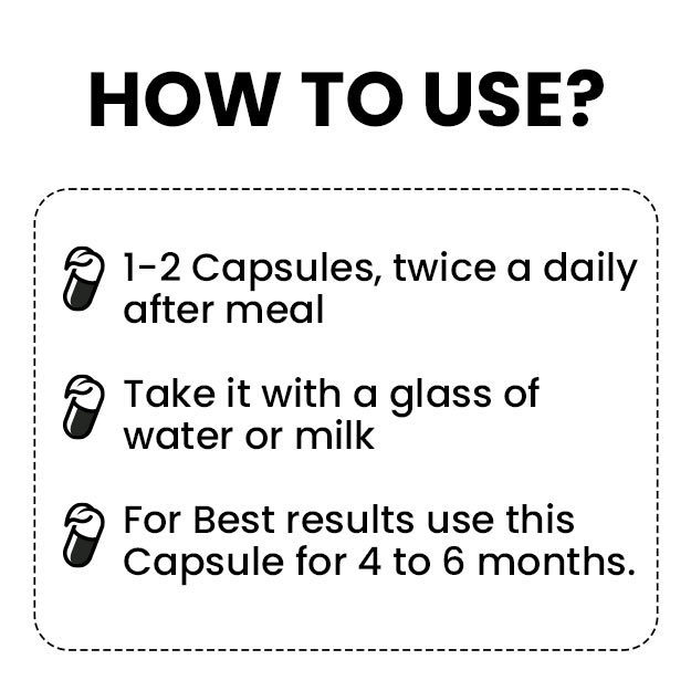
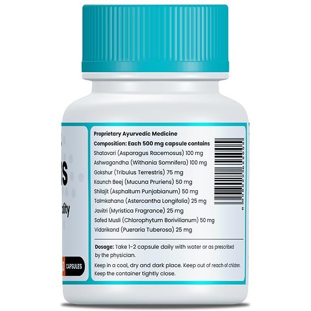
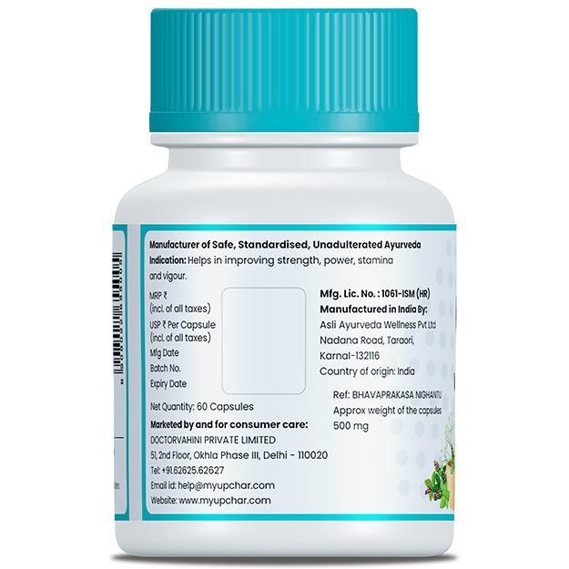
myUpchar doctors after many years of research have created myUpchar Ayurveda Urjas Capsule by using 100% original and pure herbs of Ayurveda. This ayurvedic medicine has been recommended by our doctors to lakhs of people for sex problems with good results.
Long Time Capsule
₹712  ₹799  10% OFF
BUY NOW

Following are the advantages of using a cannula:

  • By gaining intravenous access, the delivered agent enters straight into the bloodstream, thus allowing an immediate effect.
  • Intravenous access helps gain some control over the rate of administration of the agent in the best possible manner. This is more accurate than oral, intramuscular or sublingual methods.
  • IV cannulation allows delivery of desirable and suitable substances in adequate amounts in patients who cannot tolerate drugs or fluids by the oral route.
  • Some drugs cannot be absorbed by other routes; for example, drugs with a high molecular weight can only be given intravenously.
  • Once intravenous access is gained, patients do not have to experience the pain and trauma due to repetitive pricks while receiving drugs through the subcutaneous or intramuscular route.

Sometimes, drugs can be given to patients intravenously, either in a bolus form (large amounts in a short time) or by infusion over longer periods of time. Intravenous drugs can be used in the following situations:

  • Patients who suffer from serious illnesses may require drugs intravenously as there are numerous advantages of administering drugs intravenously over oral drugs. It helps in reducing mortality considerably in case of serious diseases like life-threatening bacterial infections.
  • When a drug has very low oral bioavailability (amount of drug that actually enters the bloodstream) or when a drug is only available in an intravenous form; for example, drugs like aminoglycoside antibiotics are highly polar and cannot usually be absorbed via the gastrointestinal system. Hence, they require to be delivered intravenously.
  • When patients are not able to take any medicines orally in cases of vomiting or when a doctor has advised patients to not take anything orally (i.e. nil per oral). Other routes such as rectal, sublingual, intravenous or intramuscular must be used in these patients.
  • Oral drugs may result in aspiration and choking in patients who have a low consciousness level. In such cases, intravenous routes are preferred.
  • Intravenous drugs may be used when the doctor requires the peak or highest blood level of the drug to be achieved rapidly. This can be achieved via a bolus injection as it results in a quick and identifiable increase in the amount of drug in the blood. This is essential when antibiotics are used.

A cannula should not be used in the following situations:

  • Inflammation or infection of the site where the cannula is to be inserted.
  • In patients suffering from renal failure, cannulae should not be inserted in the forearm veins as these may be needed for arteriovenous fistula formation that is required for dialysis.
  • Drugs that are considered to be irritants if injected into small veins with low-flow rates. These veins are the ones in the legs and feet.

Hence, before inserting a cannula, it is essential to understand if it is necessary for the clinical treatment of the patient or not. Sometimes, a cannula is not used immediately but it’s insertion is justified as it may be needed later on (prophylactic insertion) i.e. patients with highly serious or unstable illnesses. Most patients require intravenous cannulae for the delivery of intravenous fluids. Before a doctor decides to go ahead with this, it is important to determine if delivery of intravenous fluids is clinically appropriate and required or not.

It is important to understand that fluid and electrolyte imbalances are extremely common in patients admitted to hospitals. But, in some cases, these imbalances are not managed properly. This is especially observed in older patients. Studies suggest that this is a result of improper training of doctors who work at a junior level, as they are usually responsible for prescribing intravenous fluids in medical wards. Recent studies conducted suggested that nearly 60% of junior doctors were not formally taught the administration of intravenous fluids and nearly 40% had no idea of the patient profile or blood test reports before prescribing fluids. Junior doctors should be aware of the fact that prescribing intravenous fluids should be treated with the same importance as prescribing any drugs to the patient. Most commonly, intravenous fluids are prescribed to patients suffering from dehydration. But, even in patients of dehydration, they are not always necessary and oral fluid therapy may be sufficient for the treatment of acute gastroenteritis, especially in children. Oral fluids can even be considered in patients who are vomiting and can be given via a ryles tube when oral fluids cannot be tolerated.

It is important to know that the rate of flow in a cannula is directly proportional to the height of the reservoir of fluid and the radius of the cannula. When giving a viscous fluid like blood or for infusions given rapidly, large-sized cannulae (14-16 gauge) should be used. For less viscous fluids like crystalloids, small-sized cannulae (20-24 gauge) should be used.

Following are the types of cannulae:

  • Orange: 14 gauge cannula. It is used in operation theatres or in emergency situations such as rapid transfusion of blood, blood products or intravenous fluids.
  • Grey: 16 gauge cannula. It is also used in operation theatres or in emergency situations such as rapid transfusion of blood, blood products or intravenous fluids.
  • Green: 18 gauge cannula. It is used for patients who require blood transfusions, total parenteral nutrition and for delivering large amounts of intravenous fluids.
  • Pink: 20 gauge cannula. It is used for patients who require blood transfusions, total parenteral nutrition and for delivering large amounts of intravenous fluids.
  • Blue: 22 gauge cannula. It is used in patients when the cannula is to be inserted in small veins. It may also be used in children for delivering medications and fluids.
myUpchar doctors after many years of research have created myUpchar Ayurveda Kesh Art Hair Oil by using 100% original and pure herbs of Ayurveda. This Ayurvedic medicine has been recommended by our doctors to more than 1 lakh people for multiple hair problems (hair fall, gray hair, and dandruff) with good results.
Bhringraj Hair Oil
₹546  ₹850  35% OFF
BUY NOW

The following should be kept in mind before inserting a cannula:

  • Patients should be informed about the need for cannulation. A complete explanation of the procedure must be given to the patient before the procedure.
  • Patients should give consent for cannulation before the procedure.
  • The patient should be positioned comfortably, preferably lying down on their back.
  • Identify the non-dominant hand or arm of the patient (left or right).
  • The identified non-dominant hand or arm should be supported on a pillow or any other soft surface.
  • Before going ahead with the procedure, check if there are any other contraindications like infections, tissue damage or the need of an AV fistula (patients with renal failure).

The following equipment may be required before performing an IV cannulation: 

  • Dressing tray
  • Gloves (non-sterile)
  • Apron (non-sterile)
  • Gauze pieces or cleaning wipes
  • Intravenous cannula
  • A tourniquet (device to stop the flow of blood through a vein or artery by compressing a limb with a cord or bandage)
  • Bandages to keep the cannula in place
  • Rubbing alcohol
  • Antiseptic solution
  • Normal saline
  • Sterile syringe
  • A waste bin for sharp instruments

Following are tips to encourage filling up of suitable veins:

  • Make sure that the tourniquet is applied properly. The tourniquet should be applied to the upper arm of the patient. The tourniquet should be tight enough in terms of pressure so that it is sufficiently high to restrict venous distension but not high enough to stop the arterial flow of the blood.
  • Ask the patient to open and close the fist.
  • Make sure that the limb is situated at a position below the level of the heart.

Selecting a good vein for cannulation

A good vein should have the following qualities: 

  • It should be sufficiently bouncy
  • It should be adequately soft
  • It should be above the precious sites
  • It should easily refill after it is depressed
  • It should be easily visible
  • It should have a sufficiently large enough lumen
  • It should be straight
  • It should be well-supported
  • It should be easily palpable

Ideally, the following qualities should not be present in veins before cannulation : 

  • Veins that are thrombosed or sclerosed
  • Veins that are bruised
  • Veins that are inflamed
  • Veins that are extremely thin
  • Veins that are very fragile
  • Highly mobile veins
  • Veins that are situated near bony prominences
  • At sites of infections or previous punctures 
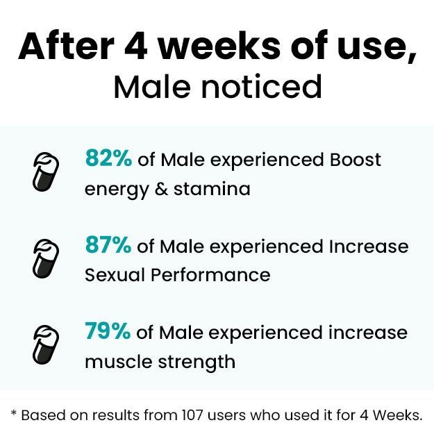
myUpchar doctors after many years of research have created myUpchar Ayurveda Urjas Energy & Power Capsule by using 100% original and pure herbs of Ayurveda. This Ayurvedic medicine has been recommended by our doctors to lakhs of people for problems like physical and sexual weakness and fatigue, with good results.
Power Capsule For Men
₹495  ₹799  38% OFF
BUY NOW

Inserting the cannula

Following are the step-by-step instructions for inserting a cannula:

  • Wash hands and prepare the equipment needed.
  • Take out the cannula from the packing and check that all the parts are functional.
  • Loosen the white cap and replace it gently.
  • The tourniquet should be applied appropriately.
  • Identify the vein to cannulate. Clean the site using an alcohol wipe and allow it to dry.
  • Non-sterile gloves should be worn.
  • The protective sleeve from the needle should be removed and the cannula should be held in your dominant hand.
  • The skin over the vein should be stretched to anchor the vein with the non-dominant hand. Make sure that you do not re-palpate the vein.
  • With the bevel side up, insert the needle at an angle of 10-30 degrees to the skin. Check for blood in the flashback chamber.
  • Slightly lower the cannula to make sure it enters the lumen of the blood vessel and does not pass through the outer wall of the vessel.
  • Now, gently push the cannula over the needle while you withdraw the guide, observing a secondary flashback.
  • The tourniquet should be released and gentle pressure should be applied.
  • Remove the white cap from the needle.
  • Remove the needle and dispose of it in the container for sharp instruments. Make sure you attach the white lock cap.
  • Apply dressing on the cannula.
  • The cannula should be flushed with a normal saline solution.

The following complications can arise because of intravenous cannulation:

  • Hematoma: Collection of blood due to failure to puncture the vein while inserting the cannula.
  • Infiltration: When the substance used enters the subcutaneous tissue and not inside the vein. It is important that the cannula is in place.
  • Embolism: Due to air or clots.
  • Inflammation: Of the vein called phlebitis.
Dr. Archimedes Amin

Dr. Archimedes Amin

Multispeciality
9 Years of Experience

Dr. Sakshi Gupta

Dr. Sakshi Gupta

Multispeciality

Dr. Himani Negi

Dr. Himani Negi

Multispeciality
9 Years of Experience

Dr. Prakashkumar P. Chandpara

Dr. Prakashkumar P. Chandpara

Multispeciality
1 Years of Experience

References

  1. B. H. Solomowitz. Intravenous cannulation: a different approach.. Anesth Prog. 1993; 40(1): 20–22. PMID: 8185086.
  2. Osti Chadani, Khadka Menuka, Wosti Deepa, Gurung Ganga, Zhao Qinghua. Knowledge and practice towards care and maintenance of peripheral intravenous cannula among nurses in Chitwan Medical College Teaching Hospital, Nepal. Nurs Open. 2019 Jul; 6(3): 1006–1012. PMID: 31367425.
  3. Rüsch Dirk, Koch Tilo, Spies Markus, Eberhart Leopold HJ. Pain During Venous Cannulation: A Randomized Controlled Study of the Efficacy of Local Anesthetics. Dtsch Arztebl Int. 2017 Sep; 114(37): 605–611. PMID: 28974291.
  4. Carr Peter J, et al. Factors associated with peripheral intravenous cannulation first-time insertion success in the emergency department. A multicentre prospective cohort analysis of patient, clinician and product characteristics. BMJ Open. 2019; 9(4): e022278. PMID: 30944127.
  5. Khadim MF, Leonard D, Moorehead RA, Hill C. Back to basics: iatrogenic intravenous cannula embolus. Ann R Coll Surg Engl. 2013 Oct; 95(7): e4–e5. PMID: 24112480.
Read on app