What is Reticulin Stain test?
Reticulin stain is commonly used in histopathology laboratories to visualise biopsy specimens of body tissues, most commonly the liver tissue but also of kidney or spleen.
Histopathological staining is employed as an aid to the treatment of diseases when a closer examination of tissues is needed to ascertain the presence of abnormalities. Staining highlights the features of the tissue and enhances its contrast, which allows the examiner to visualise it with better clarity.
Reticulin is a form of collagen, which is present in the membranes of certain body organs. Reticulin fibres form a mesh-like structure in these organs and helps support the soft tissues structures. On staining, this mesh-like structure appears in shades of black under the microscope.
Reticulin stain is made of ammoniacal silver and formalin and the process of staining is called metal impregnation. The silver initially binds to the tissue and formalin reacts with it to make a coloured precipitate that makes it possible to visualise the tissue.
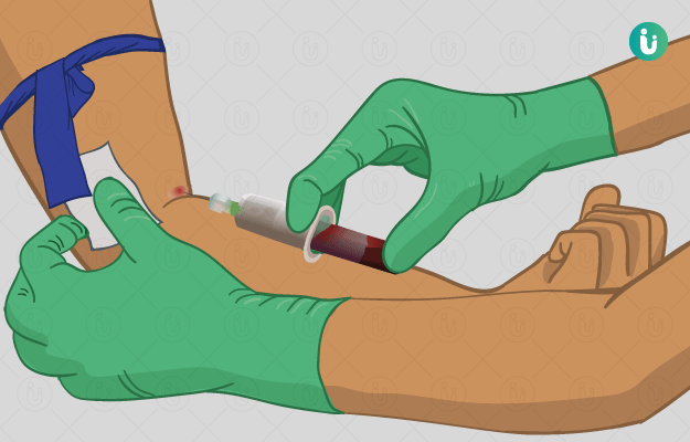
The tissue sample to be tested is obtained through biopsy.
Reticulin staining is a commonly used method for diagnosing tumours.


