Serology tests are those tests that are done to look for the presence of antigens or antibodies against specific microbes in a person’s body. The word serology comes from the word serum which is the liquid part of the blood that is left after removing fibrinogen (the clotting protein). Though serology tests are usually done on blood samples, sometimes other fluids including the urine and cerebrospinal fluid are also used for these tests.

Antigens are any foreign substance including microbes, microbial toxins, dust, and allergens. Sometimes the body starts to see its own healthy cells as antigens—this results in autoimmune diseases. Antibodies are proteins that our immune system makes to fight the antigens and eliminate them from the body. 

There are various different kinds of serology tests, depending on what is being tested (antigen or antibody) and how.

Serology tests, specifically antibody tests, are widely being used to find out asymptomatic COVID-19 cases and to look for candidates for convalescent plasma therapy—where the antibodies from a recovered patient are transferred to a person with an active infection for management of the disease.

Read on to know more about serology tests.

Read more: Who can get tested for COVID-19

  1. Types of antibodies and immune response
  2. Serology test types
  3. Why are serology tests done?
  4. How are serology results seen in a lab?
  5. Serology tests for COVID-19

  6. Serology tests limitations
Doctors for Serology

Whenever your immune system is exposed to a new pathogen (bacteria, virus, fungi, etc.), it makes specific antibodies (immunoglobulins) targeting the said microbe. The human body has five different types of antibodies—IgA, IgG, IgM, IgE, IgD. All of these antibodies are Y shaped protein complexes with two heavy chains and two light chains. All four chains have a non-changing region (Fc region) and a variable region (Fab region). The latter is what the antibodies use to identify and bind with different antigens. This variable is highly specific to antigens but some times an antibody for an antigen may cross-react with other antigens. This happens when the structure of the two antigens is similar. In other words, if the microbes that cause two different diseases are very similar, then the same antibodies could be effective against both.

IgM antibody is the first one to be produced at the time of infection. However, the IgG antibody is the most common antibody and is responsible for long-term immunity.

Antibodies stay in your blood even after the infection has cleared. A serology test can hence be used to determine if you have been exposed to the microbe—either recently or in the past. These tests can also be used to look for autoimmune diseases—conditions in which your immune system mistakenly starts to make antibodies against healthy cells in your body.

Read more: Who is immune to COVID-19 

myUpchar doctors after many years of research have created myUpchar Ayurveda Urjas Capsule by using 100% original and pure herbs of Ayurveda. This ayurvedic medicine has been recommended by our doctors to lakhs of people for sex problems with good results.
Long Time Capsule
₹712  ₹799  10% OFF
BUY NOW

Serological tests depend on antigen-antibody reactions. When an antibody identifies a particular antigen, it binds to the antigen to neutralise it and this reaction can be seen in a lab in various ways like precipitation, agglutination or colour development.

In precipitation, something can be seen settling down. In agglutination, particles can be seen clumping together. And in colour development, the test results are determined by colour change (or the absence of it).

Depending on what is being looked for (antigen or antibody) in a sample, serology tests are of two types:

  • Direct serological tests: These tests use an antiserum (serum with antibodies against an antigen or pathogen) to look for the presence of antigens in the patient’s blood/body fluids.
    So, say, you are looking for virus A in a person’s blood, then you’ll use an antiserum which has anti-A antibodies. The antigen-antibody reaction will occur if the virus A is present in the given sample.
  • Indirect serological tests: These tests look for the presence of antibodies in the patient’s serum. In this test, the patient’s serum is exposed to a certain antigen in the lab—a reaction will only occur if the patient's serum has specific antibodies against the antigen.

Serology tests are done to identify the presence of a specific antigen or antibody in the blood or body fluids of a person. These tests can be used to detect various infectious diseases, which include:

Serological tests can also be used to look for the presence of IgE antibodies, that are found in larger numbers in the blood of a person when he/she has an allergic reaction or a parasitic infection.

In the case of autoimmune diseases, specific antibodies against certain tissues of the body can be detected with serology tests. For example, an anti-dsDNA antibody test is used to detect lupus.

Anti-dsDNA antibodies are a type of autoantibodies (antibodies that attack healthy cells of the body) that target the DNA inside our cells.

myUpchar doctors after many years of research have created myUpchar Ayurveda Kesh Art Hair Oil by using 100% original and pure herbs of Ayurveda. This Ayurvedic medicine has been recommended by our doctors to more than 1 lakh people for multiple hair problems (hair fall, gray hair, and dandruff) with good results.
Bhringraj Hair Oil
₹546  ₹850  35% OFF
BUY NOW

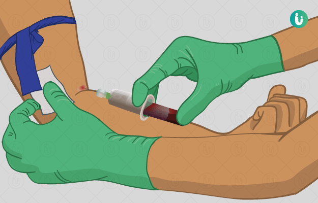
To do this test, a lab technician will first draw blood from a vein in your arm. The following are some of the techniques that are used in laboratories to visualise the antigen-antibody reaction:

  • Precipitation: In a precipitation reaction, a soluble antigen and antibody react with each other to form a precipitate that can be easily visualised with the naked eye. In this method, a deposit or precipitate will only be seen if the test is positive.
    Precipitation reactions can be done in a solution (fluid phase) or a gel phase. In the former, fluid solution of an antigen is layered on the antibody solution (or the other way round)—depending on whether the patient's blood sera is being tested for the presence of antigens or antibodies, it will take the place of one of these layers.
    The two layers will mix and the antibodies and antigens (if they are both present) will travel towards each other through passive diffusion. The precipitate, if any, will be visible somewhere in the middle of the two solutions, wherever the antigens and antibodies are present in equal amount (zone of equivalence). No precipitate will be visible in the area of antigen or antibody excess, which would be above and below the precipitate line.
    The gel phase precipitation is also a passive diffusion method. In this, agarose gel is let to sit in a Petri plate and wells are cut out in the gel. Now, in the central well, an antigen or antibody solution is added and the opposite solution is added to the surrounding wells. The solutions will travel through the gel and lines of precipitation will be formed at the zone of equivalence.
    In another type of gel phase precipitation, antiserum is added into the gel before it solidifies and antigen solution is put into the dug wells. Precipitation occurs around the wells in the zone of equivalence. The latter method is called radial immunodiffusion and the former is called the Ouchterlony technique.
    Another kind of precipitation is electrodiffusion. In this, the movement of antigen and antibody molecules occurs under the effect of an electric current.
  • Agglutination reaction: In this technique, either antigens or antibodies are bound to a particle such as latex beads, charcoal particles or red blood cells and then the antigen-antibody reaction is visualised for the formation of visible clumps—agglutination reaction.
    For example, in an agglutination reaction that uses latex beads and to identify antibodies in a person, the antigen would be bound to the latex beads and then it will be exposed to the patient’s serum. Visible clumping will occur if the given sample has antibodies against the test antigen.
    For the agglutination reaction, whole bacteria (or other pathogens) may also be used as the antigen. It is then called direct agglutination. The antibodies (if present) in the patient’s serum will attack the bacteria and form clumps.
    Agglutination reactions are more sensitive than precipitation reactions. The sensitivity of a test refers to its ability to correctly identify people who have the particular infection being tested for—basically, the test gives more true positives and fewer false negatives.
  • Immunoassays: Immunoassays use antigen-antibody reactions to quantify the amount of antigen or antibody present in a given sample. The following are some types of immunoassays:
    • ELISA: Enzyme-Linked Immunosorbent Assay (ELISA) uses an enzyme to generate colour when an antigen-antibody reaction occurs. This enzyme is either linked to the antigen or antibody, depending on what needs to be checked for in the unknown sample.
      Some enzymes used for ELISA include alkaline phosphatase and horseradish peroxidase.
      Here is how an ELISA test is done to look for an antibody: A solid-phase such as a microtiter plate is used, to which the specific antigen (against which the antibody needs to be checked for) is attached. Then the patient’s serum is added into the well. If the serum has antibodies against the antigen in the well, the antigen-antibody reaction will occur. Any free antibody will be washed out of the plate by using a specific solution. Now, an enzyme-linked antibody will be added to the plate. This antibody is specific to the antibody that needs to be found in the given sample. If the first antibody is in the plate, this second antibody will bind to it. All the unbound secondary antibodies will also be washed. Next, the substrate for the enzyme will be added to the solution. The enzyme will react with the substrate and show up colour if any secondary antibody is left in the plate. By specific instruments, the colour intensity is checked and the level of antibodies will be obtained.
    • Western blotting: Western blot is yet another type of immunoassay that can be used to identify specific proteins in a given sample. In this technique, proteins/antigens from a solution are first separated onto a polyacrylamide gel under the influence of electric current. From this gel, the antigens are transferred to a filter paper, which like the ELISA microtiter plates, becomes the solid surface. Next, the filter paper is exposed to enzyme-linked antibodies. And finally, the substrate of the enzyme is added to the solution. If the antibodies have bound to the antigen and are not washed out, the enzyme would react with the substrate and produce colour. The intensity of the colour would tell how much of the target protein was present in the solution.
    • Immunofluorescence: Immunofluorescence is most commonly used to look for specific antigens in a sample. In this technique, antibodies are labelled with specific fluorescent dyes. These antibodies will bind to the antigen (if present) in a sample. Then the antigen-antibody complexes will be exposed to light of a certain wavelength, upon which, they will show fluorescence, which can be visualised as coloured light. For example, fluorescein, a dye, when exposed to blue light, emits yellow-green fluorescence. This method does not quantify antigens. However, a more modern and automated method of immunofluorescence uses a chamber called fluorescence-activated cell sorter. When the fluorescence labelled antigen-antibody complexes are passed through this machine, it flashed laser beams on it and counts every single cell that is labelled with the fluorescence.

Currently, serological testing for COVID-19 is being done to look for the presence of antibodies against the SARS-CoV-2 coronavirus in a person’s blood. SARS-CoV-2 or severe acute respiratory syndrome coronavirus 2 is the pathogen which causes COVID-19.

You may be asked to get an antibody test done if:

  • You are a convalescing (recovering) patient and are a potential donor for antibody-rich plasma.
  • If your physician thinks that you have been exposed to the virus in the past and may have developed the disease but were not tested then.
  • If you get a negative result in the RT-PCR test and your physician still wants to confirm if you have had the infection.

If you were not tested positive for COVID-19 in the past and still show the presence of antibodies, it means you have been exposed to the virus already. Though, it is still unknown whether a person becomes immune to the virus once they develop antibodies.

Types of antibody tests: According to the US Centers for Disease Control and Prevention, there are two types of antibody tests—those that look for binding antibodies and those that look for neutralising antibodies. Here is what these tests entail:

  • Binding antibody tests: Binding antibodies are those that bind to the virus or the antigen but do not necessarily neutralise the virus. To look for binding antibodies, the purified proteins of the SARS-CoV-2 virus are used. (In terms of structures, SARS-CoV-2 comprises RNA as genetic material encased in a shell with spike proteins on the surface—these spike proteins are the source of purified proteins used in this test.)
    Since these tests do not use the live virus, they can be done in labs with lower biosafety levels.
    Most commonly ELISA test for COVID-19 is done to look for COVID-19 antibodies. However, some point of care tests are also used for antibody detection. The latter is based on lateral flow assays (a pregnancy test is a type of point of care test) and can be done using a small drop of the patient’s blood.
  • Neutralising antibody tests: Neutralising antibodies are those that actually bind to the virus and neutralise it. So far, the US Food and Drug Administration (FDA) has not approved any neutralising antibody test for COVID-19. These tests use live virus and hence need special labs to avoid infection to the people working in the lab.
    Plaque reduction neutralisation tests (PRNT) are most commonly used to check for the presence of neutralising antibodies. In this test, a fixed amount of virus is mixed with the patient’s serum and the solution is put over a microtiter plate that contains susceptible cells. Then, the effects of antibodies are measured by noting the size and number of plaques made in the plate. In a microtiter plate, plaques are clear areas where the virus has infected the cells and produced progenies. Usually, the serum sample is serially diluted before adding it to various tubes with a specific amount of virus. This helps in clearly identifying plaque areas and the number of plaques—if there is an inadequate number of antibodies, the plaques may merge and would be difficult to count.

Types of antibodies COVID-19 antibody tests look for: The tests specifically look for the presence of IgG or IgM antibodies against the virus antigens. IgA antibodies are looked for to determine mucosal immunity. These antibodies are found in blood and mucosal surfaces like the nose. However, the importance of this antibody in COVID-19 has not been determined so far.

SARS-CoV-2 antigens: So far, the spike protein of the SARS-CoV-2 virus, the receptor-binding domain of the virus and the outer protein coat have been used as antigens. The spike protein is what the COVID-19 causing virus uses to bind to and enter into healthy cells. The receptor-binding domain is the specific site on the spike protein that binds to the ACE2 receptors on the surface of host cells. (Read more: What are ACE2 receptors and what do they have to do with COVID-19?)

Antibody test results: Here is what the antibody test results mean:

  • Presence of IgM antibodies usually denotes the presence of recent infection and that the person’s immune system is actively fighting against the virus still. On the other hand, the presence of IgG antibodies means that it has been a while the person had the infection, more tests would reveal if he/she is still infectious or not.
    Positive result: If both IgG and IgM antibodies are found in the blood of a person, it means the infection is active and likely began about 14 days back. In such cases, the person is still infectious and needs to be isolated. In case the test comes back positive for IgM antibodies only, the person is suggested to get a repeat test within 14 days or so of the infection, to see if their body is producing IgG antibodies yet.
    Negative result: If a person tests negative for both the antibodies, it could either mean that he/she has not been exposed to the virus before or his/her immune system has not started making antibodies against the virus yet.
myUpchar doctors after many years of research have created myUpchar Ayurveda Urjas Energy & Power Capsule by using 100% original and pure herbs of Ayurveda. This Ayurvedic medicine has been recommended by our doctors to lakhs of people for problems like physical and sexual weakness and fatigue, with good results.
Power Capsule For Men
₹799  ₹799  0% OFF
BUY NOW

Even though serological tests give both qualitative and quantitative results—they tell you whether the antigens/antibodies are present and in what amounts—and are effective in checking the immune response of a person, these tests have the following limitations:

  • Our immune system produces various kinds of antibodies (polyclonal response) against an infection. So, person A may produce one type of antibody clones more than person B but still have an effective immune response. This increases the uncertainty of the test.
  • Also, different strains of a microbe may change the serology of the said microbe—what kind of antibodies they bind to and how strongly.
  • It takes a while before antibodies form in a person’s body. So, antibody tests cannot be used to diagnose a disease.
Dr Rahul Gam

Dr Rahul Gam

Infectious Disease
8 Years of Experience

Dr. Arun R

Dr. Arun R

Infectious Disease
5 Years of Experience

Dr. Neha Gupta

Dr. Neha Gupta

Infectious Disease
16 Years of Experience

Dr. Anupama Kumar

Dr. Anupama Kumar

Infectious Disease

References

  1. Jacofsky David, Jacofsky Emilia M., Jacofsky Marc. Understanding Antibody Testing for COVID-19. J Arthroplasty. 2020 Apr 27. doi: 10.1016/j.arth.2020.04.055 [Epub ahead of print]. PMID: 32389405.
  2. AIDS Info [Internet]. National Institute of Health. U.S. Department of Health and Human Services. Maryland. US; Serologic Test
  3. Michigan Medicine: University of Michigan [internet]. US; Immunoglobulins
  4. Cochrane UK [Internet]. The Cochrane Collaboration. London. UK; Sensitivity and specificity explained: A Cochrane UK Trainees blog
  5. University of Hawaii [Internet]. Hawaii. US; Chapter 5 Antigen-Antibody Interactions, Immune Assays and Experimental Systems
  6. UCLA health [Internet]. University of California. Oakland. California. US; What is Antibody (Serology) Testing
  7. Science Direct (Elsevier) [Internet]; Binding antibody
  8. World Health Organization [Internet]. Geneva (SUI): World Health Organization; Guidelines for Plaque reduction neutralization testing of human antibodies to dengue viruses
  9. US Food and Drug Administration (FDA) [internet]. Maryland. US; EUA Authorized Serology Test Performance
  10. Centers for Disease Control and Prevention [internet]. Atlanta (GA): US Department of Health and Human Services; Interim Guidelines for COVID-19 Antibody Testing
  11. American Society for Microbiology [Internet]. Washington DC. US; COVID-19 Serology Testing Explained
  12. Fierz W. Basic problems of serological laboratory diagnosis. Methods Mol Med. 2004;94:393‐427. PMID: 14959841.
  13. Jenni Punt, Sharon Stranford, Patricia Jones, Judith A Owen. Kuby Immunology. 8th eds. WH Freeman: Macmillan Publishers.
Read on app
cross
Ask your health query from live doctors now!