What is a pelvis MRI?
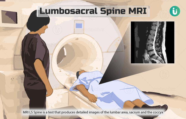
An MRI—magnetic resonance imaging—is a scan that produces images of the inside of the body with the help of magnets and radio waves. These images enable the healthcare provider to observe tissues like different organs and muscles in the body without any obstruction from bones.
The pelvis MRI captures images of the organs and tissues between the hip bones—the pelvic area. The structures found in the pelvic region include the large intestine, small intestine, urinary bladder, lymph nodes, pelvic bone and the male or female reproductive organs.
Pelvis MRI helps in diagnosing problems like pain in the hips and the spread of cancer in the pelvic area. An MRI is considered safer than x-rays and CT scans as it does not use radiation.
Each MRI image is known as a slice. Dozens to hundreds of slices are produced in each test. These slices may be printed immediately or stored on the computer.
(Read more: Pelvic ultrasound)


