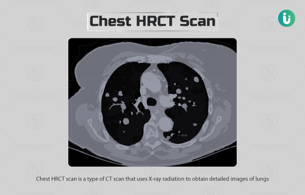
Chest HRCT (High-resolution computed tomography) scan, also known as HRCT of the lungs, is a type of CT scan that uses X-ray radiation to obtain detailed images of lungs.
Both CT scan and HRCT scan take thin-slice (cross-sectional) images of the chest to produce images; however, the slices are much thinner in case of an HRCT scan. Thus, the images provided by an HRCT scan have a much higher resolution than a normal CT scan. These images are ideal for the evaluation of a group of lung disorders called interstitial lung disease (also known as diffuse interstitial lung disease or diffuse lung disease) in which the tissue (interstitium) that supports the air sacs of the lungs becomes inflamed and stiff.
A full HRCT scan protocol usually includes the following:
- Expiratory images: This scan is performed with the person lying face up and images are obtained at the end of expiration (when the person breathes out).
- Prone images: This scan is performed with the person lying face down and images are obtained at full inspiration (when the person takes a deep breath).

