What is ankle joint magnetic resonance imaging (MRI)? 

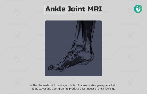
MRI of the ankle joint is a diagnostic test that uses a strong magnetic field, radio waves and a computer to produce clear images of the ankle joint.

The ankle joint connects the bones of our leg to the bones of our foot and allows us to move our foot up and down. It is reinforced by different soft tissues, such as:

  • Ligaments: Strong tissues that hold the bones together.
  • Tendons: Tissues that attach muscles to the bones.
  • Cartilage: Smooth tissue that covers the ends of the bones.
  • Synovium: A thin layer that lines the space within the joint.

Ankle joint MRI helps to detect injuries and abnormalities in the soft tissues and bones of the ankle. It is the preferred method to obtain good soft tissue images.

Your doctor may or may not use a contrast dye to perform this test. A contrast dye helps see images in better clarity (due to the contrast created) and is usually done in cases such as fractures, or cartilage or muscle tear. 

  1. Who cannot have ankle joint MRI?
  2. Why is an ankle joint MRI done?
  3. How should I prepare for an ankle joint MRI?
  4. How is an ankle joint MRI done?
  5. How will an ankle joint MRI feel like?
  6. What do the results of an ankle joint MRI mean?
  7. What are the risks and benefits of an ankle joint MRI?
  8. What happens after an ankle joint MRI?
  9. Contrast vs Non-contrast ankle joint MRI
  10. What are the other tests that can be done with an ankle joint MRI?

Because of the strong magnetic field used for the test, MRI is avoided if you have the following implanted medical devices:

  • Cardiac pacemakers
  • Cardiac defibrillators 
  • Cochlear (ear) implants 
  • Clips for brain aneurysms
  • Metal coils within blood vessels

 MRI is also avoided in the first trimester of pregnancy.

Your healthcare practitioner may order this test if you have symptoms of an ankle injury, especially if they last for more than six weeks and are not clear by other tests like an x-ray. These symptoms include:

  • Pain or soreness
  • Decreased motion of the ankle joint 
  • Bruising
  • Swelling or redness
  • Instability of the ankle
  • Difficulty in walking

An ankle joint MRI may also be performed in the following situations:

  • To check for birth defects in the ankle 
  • To detect broken bones (fractures)
  • To find the cause of ankle pain that does not improve with treatment

 The following preparations are needed for the ankle joint MRI:

  • Your doctor may ask you to fill out a screening form before the test.
  • You may have to fast for four to six hours before the test.
  • If you are afraid of enclosed spaces, the doctor may prescribe a sedative. 
  • You will be asked to wear loose and comfortable clothes without metal fasteners or a hospital gown for the test.
  • Avoid carrying any metal items inside the examining room, including:
    • Jewellery, credit cards and watches, as they can get damaged
    • Pens, eyeglasses and pocket knives, as they may fly across the room
    • Pins, hairpins and metal zippers, as they may blur the images

The following steps are performed for an ankle joint MRI scan:

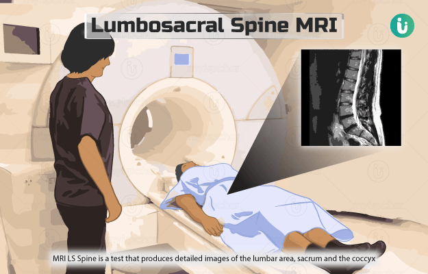
  • You will be asked to lie down on your back on the motorised scan table.
  • You will be positioned feet first such that when the table slides into the wide circular tunnel of the MRI machine, your head will remain outside and your feet will be in the centre of the tunnel.
  • If a contrast MRI scan is done, the technologist may inject a contrast dye either in a vein of your arm or into the affected joint.
  • He/she will place an extra camera over the affected ankle.
  • You will be able to hear some noises from the machine when the images are captured. 
  • Make sure to remain still while the test is being done as movements can obscure the MRI images.

The ankle joint MRI takes 30 to 35 minutes.

Joint Capsule
₹716  ₹799  10% OFF
BUY NOW

MRI scan is not painful. However, you may have slight discomfort from remaining still for the procedure. If you feel cold inside the room, the technologist may provide you with a blanket. The machine can be noisy, but you will be provided with ear protection.

Ankle joint MRI can help diagnose the following conditions:

  • Synovitis (inflammation of the synovium)
  • Tenosynovitis (inflammation of the protective sheath of a tendon)
  • Tear or rupture of the Achilles tendon in the ankle
  • Ankle impingement (abnormal entrapment of tissues that causes pain)
  • Fracture
  • Degeneration due to age
  • Cartilage injuries
  • Bone tumour
  • Arthritis
  • Ligament injuries
  • Muscle damage
  • Contusion (an injury to the skin and underlying tissues of the ankle)
  • Avascular necrosis (a condition with bone tissue death due to insufficient blood supply to the bone)

The benefits of an MRI scan are:

  • It is a painless and non-invasive method. 
  • It provides detailed soft tissue images.
  • There are no radiations involved in this test. 
  • It can diagnose conditions that are hidden by bones in other imaging tests.
  • The dye used in MRI shows lesser allergic reactions as compared to iodine-based dyes used in x-ray and computed tomography (CT) scan

The risks of an MRI scan are:

  • The magnetic field used may damage any implanted medical devices. 
  • Rarely, an allergic reaction to gadolinium may be seen.
  • In individuals with serious kidney disease, the injection of gadolinium contrast may cause nephrogenic systemic fibrosis, a condition in which there is excessive fibrous connective tissue formation (fibrosis) in the skin and internal organs.

You can continue with your normal diet and activities after the test. If a sedative is given, you may be asked to bring someone who can drive you back home.

The doctor may administer a gadolinium contrast dye in the arm or in the affected joint to improve the clarity of the images of the MRI scan if needed.

Some conditions that require the injection of a contrast dye are:

However, you may not always need a contrast dye injection. Some conditions that do not require the injection of a contrast dye are as follows:

  • Arthritis
  • Fracture
  • Muscle/tendon tear
  • Cartilage tear
  • Ligament tear
  • Joint pain

If you have serious kidney disease, the contrast may cause nephrogenic systemic fibrosis. In rare cases, you may be allergic to the contrast and will need to inform the doctor.

An arthroscopy may be performed with an MRI to confirm ankle fractures.

Disclaimer: All results must be clinically correlated with the patient’s complaints to make a complete and accurate diagnosis. The above information is provided from a purely educational point of view and is in no way a substitute for medical advice by a qualified doctor.

References

  1. Radiological Society of North America (RSNA) [internet]. Oak Brook. Illinois. USA; Magnetic Resonance Imaging (MRI) Safety
  2. Cedars Senai [Internet]. California. US; MRI Lower Extremities (Leg)
  3. Arthritis Foundation [Internet]. Georgia. Australia; Ankle Anatomy
  4. Wolfe MW, et al. Management of Ankle Sprains. Am Fam Physician. 2001 Jan 1;63(1):93-105
  5. Thomsen HS, Reimer P. Intravascular contrast media for radiography, CT, MRI and ultrasound. In: Adam A, Dixon AK, Gillard JH, Schaefer-Prokop CM, eds. Grainger & Allison's Diagnostic Radiology: A Textbook of Medical Imaging. 6th ed. Philadelphia, PA: Elsevier Churchill Livingstone; 2015:chap 2
  6. Kosmas C, Schreibman KL, Robbin MR. Foot and ankle. In: Haaga JR, Boll DT, eds. CT and MRI of the Whole Body. 6th ed. Philadelphia, PA: Elsevier; 2017:chap 64
  7. Hogrefe C, Jones EM. Tendinopathy and bursitis. In: Walls RM, Hockberger RS, Gausche-Hill M, eds. Rosen's Emergency Medicine: Concepts and Clinical Practice. 9th ed. Philadelphia, PA: Elsevier; 2018:chap 107
  8. Harvard Libarary [Internet]. Harvard University. Massachusetts. US; Ankle impingement
  9. Biundo JJ. Bursitis, tendinitis, and other periarticular disorders and sports medicine. In: Goldman L, Schafer AI, eds. Goldman-Cecil Medicine. 25th ed. Philadelphia, PA: Elsevier Saunders; 2016:chap 263
  10. Cannon DL. Hand infections. In: Azar FM, Beaty JH, Canale ST, eds. Campbell's Operative Orthopaedics. 13th ed. Philadelphia, PA: Elsevier; 2017:chap 78
  11. Rosenberg Zehava S., Beltran Javier, Bencardino Jenny T. MR Imaging of the Ankle and Foot. RadioGraphics. 20(1).
  12. Nemours Children’s Health System [Internet]. Jacksonville (FL): The Nemours Foundation; c2017; A to Z: Contusion (Bruise), Ankle
  13. Cleveland Clinic [Internet]. Ohio. US; Osteonecrosis (ON)
  14. Hayeri MR, et al. Soft-Tissue Infections and Their Imaging Mimics: From Cellulitis to Necrotizing Fasciitis. Radiographics. 2016 Oct; 36(6): 1888-1910. PMID: 27726741.
Read on app
cross
Ask your health query from live doctors now!