Aortic dissection

Dr. Suvansh Raj NirulaMBBS

November 17, 2020

January 04, 2021

Aortic dissection
Aortic dissection

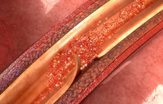
An aortic dissection is a disruption in the wall of the aorta due to intramural bleeding. This dissection results in the separation of the layers of the aortic wall, to form a true and false blood column or lumen.

The aorta is the largest blood vessel in the human body. This artery performs the vital function of carrying blood from the heart, and through the chest and the abdomen. The

arteries that branch off from the aorta supply all the major parts of the body.

Uncontrolled hypertension, cholesterol plaque build-up in the arteries (atherosclerosis), an aortic aneurysm (the blood vessel becomes weak and bulges in places), a heart valve defect and birth defect (aortic coarctation) are some of the risk factors for an aortic dissection.

An aortic dissection is one of the most commonly occurring emergencies affecting the aorta. An aortic dissection is more likely to occur in males as compared to females (nearly 70% of cases of aortic dissection are seen in men). The age group that is most affected is 50-70 years.

In terms of types, an aortic dissection can be acute, subacute or chronic.

1) Acute aortic dissection: Less than 14 days since the disease onset

2) Subacute aortic dissection: 15-90 days since the disease onset

3) Chronic aortic dissection: More than 90 days since the onset of the aortic dissection.

Read on to know more about aortic dissection, what causes it, its symptoms, how it can affect the patient’s health, its diagnosis, treatment and prevention.

Types of aortic dissection

There are two different classifications that are universally accepted. They are as follows:

Stanford classification

  • Type A involves the ascending section of the aorta
  • Type B does not involve the ascending section of the aorta

DeBakey classification

  • Type 1: Involves the aorta, aortic arch and the descending aorta
  • Type 2: Involves only the ascending aorta
  • Type 3: Involves the descending aorta distal to the left subclavian artery
myUpchar doctors after many years of research have created myUpchar Ayurveda Urjas Capsule by using 100% original and pure herbs of Ayurveda. This ayurvedic medicine has been recommended by our doctors to lakhs of people for sex problems with good results.
Long Time Capsule
₹712  ₹799  10% OFF
BUY NOW

Symptoms of aortic dissection

A patient suffering from aortic dissection can present with the following signs and symptoms:

  • Most common patient profile is an elderly hypertensive man (usually more than 60 years old)
  • with pain in his chest which starts suddenly.
  • Patients usually describe the chest pain as extremely severe, maximal at the start and is “ripping”, “sharp” or “tearing” in nature.
  • Sometimes the pain may radiate to different areas such as the back or groin of the patient.

Some symptoms can present due to an occlusion of various small arteries caused during the course of the dissection. They can be:

  • Angina due to occlusion of the coronary arteries i.e. arteries supplying the heart muscles (chest pain or shortness of breath due to limited blood supply to the heart)
  • Restricted blood supply to the limbs
  • Neurological symptoms due to the involvement of carotid arteries (major arteries in the head and neck region)
  • On examination, sometimes a difference in blood pressure may be observed in the left and right limbs

Differential diagnosis for aortic dissection

While diagnosing aortic dissection, it is extremely important to rule out other important causes of chest pain as other causes if untreated can prove to be fatal and the treatment of each condition varies from one cause to the other. The most important causes of chest pain to consider are as follows:

  • Acute coronary syndrome which mainly includes myocardial infarction or heart attack (death of heart muscles due to loss of blood supply to different regions of the heart)
  • Aortic valvular defects such as aortic regurgitation
  • Aortic aneurysms (ballooning of the aorta)
  • Pericarditis (Inflammation of the thin layer surrounding the heart)
  • Mediastinal tumors (Benign or malignant). Mediastinum is defined as the space between the two lungs in the chest cavity.
  • Musculoskeletal causes of chest pain
  • Pulmonary embolism (a blood clot occluding the pulmonary arteries i.e. the arteries that carry blood deprived of oxygen from the right side of the heart to the lungs)
  • Cholecystitis (Inflammation of the gall bladder) that can also sometimes present with pain radiating to the back

Out of all these differentials, myocardial infarction is the most important one to rule out which can be present alongside an aortic dissection.

Aortic dissection tests and diagnosis

The diagnosis of an aortic dissection should be made on the basis of the patient profile, clinical features and radiological investigations. The ultimate goal is to make a definitive diagnosis and successfully differentiate aortic dissection from a myocardial infarction. Sometimes, both these conditions may co-exist.

The following investigations should be performed:

  • Electrocardiogram: The goal is to look for signs of myocardial infarction on the electrocardiogram strip. Sometimes, patients with Stanford classification type A may have electrocardiogram changes similar to myocardial infarction.
  • Radiological investigations:
    • Chest X-ray: A chest X-ray can sometimes rule out other important causes of chest pain.
    • An aortic dissection cannot be ruled out just on the basis of an X-ray.
    • Transthoracic or transesophageal ultrasound – This is the most appropriate investigation to be performed in patients who are critically ill (Hemodynamically unstable i.e. low blood pressure and a raised pulse rate). A transthoracic or transesophageal ultrasound can localize the site and help in successfully classifying an aortic dissection based on the Stanford or DeBakey system.
    • Computed tomography aortography – This procedure involves taking X-rays of the aorta after the injection of a contrast material into the aorta via a catheter (a flexible tube). It is the most appropriate investigation for a hemodynamically stable patient.
    • Magnetic resonance imaging scans (MRI scans) – Even though Computed tomography aortography is still considered to be the investigation of choice, Magnetic resonance imaging scans offer a higher sensitivity (98%) and specificity (98%). MRI scans work in a similar way as a CT scan but, they use magnetic waves instead of X-rays.
    • Colour flow doppler: It is an additional investigation that helps in the detection of an aortic valve problem like an aortic regurgitation.
    • Intravascular ultrasound scans: They can be useful in detecting the involvement of side branches of the aorta. These may include the renal arteries, the mesenteric arteries and the coeliac arteries.

As per recent studies and accuracy testing, Magnetic resonance imaging angiography may soon replace a conventional angiography to diagnose aortic dissection.

myUpchar doctors after many years of research have created myUpchar Ayurveda Kesh Art Hair Oil by using 100% original and pure herbs of Ayurveda. This Ayurvedic medicine has been recommended by our doctors to more than 1 lakh people for multiple hair problems (hair fall, gray hair, and dandruff) with good results.
Bhringraj Hair Oil
₹546  ₹850  35% OFF
BUY NOW

Aortic dissection patient care and treatment

A patient diagnosed with aortic dissection may be managed by conservative measures with or without surgery depending on the type of aortic dissection that is classified as per the Stanford or DeBakey classification systems. General measures that should be taken for all patients regardless of definitive management. They are as follows:

  • Establish intravenous access in all the patients
  • Reduction of severe pain using analgesic drugs such as morphine
  • All patients diagnosed with aortic dissection should be managed in the intensive care unit (ICU) or a high dependency unit (HDU)
  • The blood pressure of the patient must be monitored and brought under control as soon as possible to prevent further complications. The target systolic blood pressure is between 100-120 mm Hg.
  • The most commonly used antihypertensive medication are beta-blockers such as propranolol.
  • These drugs are given via an intravenous route. Beta-blockers are a class of drugs that block the stimulation of adrenergic receptors responsible for reducing cardiac action.
  • The patient may display signs of blood loss due to the effect of double blood column formation.
  • Therefore, separate Intravenous lines are needed for the infusion of blood and other drugs.

Surgery for aortic dissection

Surgical intervention works by placing a stent or a graft in the aorta. The requirement of surgery depends on the type of aortic dissection as per the universal classification systems.
  • Stanford type A (DeBakey type 1 and 2) aortic dissection – In these patients, the goal of the surgical procedure is to reduce the frequency of aortic rupture, resolving aortic valvular diseases like aortic regurgitation and to prevent complications like pericardial tamponade (compression of the heart due to fluid buildup in the sac surrounding the heart). A graft is placed in the ascending aorta. Replacement of the aortic arch may also be performed.
  • Stanford type B (DeBakey type 3) aortic dissection – These patients may undergo a thoracic endovascular aortic repair (TEVAR). Recent studies suggest that this procedure has better five-year survival and slows down the progression of the disease. In some patients in whom TEVAR is not applicable can undergo an open surgical repair.

Chronic aortic dissection surgery

Patients suffering from chronic aortic dissection of the descending aorta may only require continuous medical treatment with frequent follow-ups using ultrasound scans. Surgery in patients of a chronic aortic dissection may result in conditions like paraplegia (impaired motor and sensory functions of the lower limbs). If repair of the dissection is required, then it can be performed via open surgical or endovascular approach.

Prognosis or outcome in aortic dissection

Aortic dissection usually offers a poor prognosis and is associated with a high mortality rate. Nearly 20% of people usually die before reaching the hospital. In patients with complicated aortic dissection is associated with a mortality rate of 80%.
Early conservative measures with adequate blood pressure control according to the target range drastically improves the outcome of the disease. After successful surgery may improve the chances of survival, still there is still a high postoperative and perioperative mortality associated with these patients.

Patients in whom late deaths are seen (after a significant duration from the onset of disease) are usually associated with aortic rupture.

myUpchar doctors after many years of research have created myUpchar Ayurveda Urjas Energy & Power Capsule by using 100% original and pure herbs of Ayurveda. This Ayurvedic medicine has been recommended by our doctors to lakhs of people for problems like physical and sexual weakness and fatigue, with good results.

Power Capsule For Men
₹719  ₹799  10% OFF
BUY NOW

Complications of aortic dissection

The complications that may present in these patients depends on the location of aortic dissection. The complications that can be associated with aortic dissection are as follows:

  • Low blood pressure (hypotension): Low blood pressure in aortic dissection is suggestive of patients with cardiac tamponade (compression of heart due to accumulation of fluid in the sac around the heart) or a myocardial infarction/heart attack.
  • Aortic valvular defects such as aortic regurgitation.
  • Heart failure
  • Fever due to the release of pyrogens (substances which are released into the blood that can result in fever) from the aortic wall
  • Coeliac artery involvement – Presents with severe continuous abdominal pain along with abnormalities on various blood tests.
  • Obstruction of the superior vena cava / Superior vena cava syndrome (Vessel from which carries deoxygenated blood (deprived of oxygen) from various parts of the body to the right side of the heart.
  • Poor perfusion of the mesentery (an organ attached to the intestines that allows blood vessels to supply the intestines) results in poor disease outcome.
  • Involvement of the distal aorta is sometimes associated with the involvement of renal arteries (arteries that arise from the abdominal aorta and supply blood to the kidneys). In such patients, immediate surgery is to be considered.

Prevention of aortic dissection

Some people are more prone to developing aortic dissection. These patients are usually suffering from various genetically inherited diseases such as Marfan’s syndrome and Ehler-Danlos syndrome. Preventive measures must be taken to prevent the occurrence of aortic dissection in these patients. They include the following:

  • Lifelong medications: Beta-blockers
  • Preventive surgical procedures such as replacement of the aortic root in case it is dilated
  • Frequent monitoring of the aortic diameter
  • Minor reduction in physical exertion and exercise