What is Upper GI Endoscopy? 

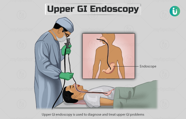
Upper GI endoscopy, also referred to as gastroscopy or oesophagogastroduodenoscopy, is used to diagnose and treat upper GI problems. It helps your doctor to view the inside of your upper GI (or digestive) tract. The upper GI tract consists of the food pipe (oesophagus), stomach and the first part of the small intestine (duodenum). 

This test is performed using an endoscope, which is a long, flexible tube with a tiny camera and a light attached at one end. The tube is inserted through the mouth into the oesophagus, stomach and duodenum. The video images captured by the camera can be viewed on a screen in real-time.

Read more: Why is an endoscopy done?

  1. Why is an Upper GI Endoscopy done?
  2. Who cannot have an Upper GI Endoscopy?
  3. How should I prepare for an Upper GI Endoscopy?
  4. What is the procedure for an Upper GI Endoscopy?
  5. How will an Upper GI Endoscopy feel?
  6. What do the results of an Upper GI Endoscopy mean?
  7. What are the risks and benefits of an Upper GI Endoscopy?
  8. What happens after an Upper GI Endoscopy?
  9. What are the other tests that can be done with an Upper GI Endoscopy?

Your doctor may perform this test if you have symptoms such as:

Troubled by obesity? Failed to lose weight? Now control weight easily by myUpchar Ayurveda Medarodha Weight Control Tablet. Get started today and take steps towards a healthy life.

This test is also used to:

  • Stop bleeding in the upper GI tract 
  • Perform endoscopic surgery, laser therapy or widen a narrowed area
  • Take tissue samples for testing (biopsy
  • Remove food or other items stuck in the upper GI tract 
  • Treat swollen veins in the oesophagus (oesophageal varices)
  • Remove cancerous tumours or non-cancerous growths (polyps)
  • Place a feeding tube or drainage tube in the GI tract
Digestive Tablets
₹312  ₹349  10% OFF
BUY NOW

Upper GI endoscopy is contraindicated in people with the following conditions:

  • Swelling of the inner wall of the abdomen (peritonitis)
  • A tear in the wall of the intestine (perforated bowel)
  • Severe inflammation and bulging of the intestine (toxic megacolon)
  • Bleeding disorders
  • Bulging of a major blood vessel in the abdomen (aneurysm of the abdominal aorta)
  • Very low count of a type of white blood cell (severe neutropenia)

Your healthcare provider will ask you to sign a consent form that gives him permission to conduct this procedure. They will usually ask you to stop eating for six to eight hours and avoid drinking for two to three hours before the procedure. You should inform your doctor if you:

  • Are allergic to any medications (including anaesthesia medicines), latex or tape
  • Are pregnant 
  • Have a bleeding disorder 
  • Are taking any blood-thinning medicine, aspirin or ibuprofen

You may be asked to avoid some medicines before the test. Your doctor may ask you to take a laxative, an enema or a special liquid that helps prepare your bowel for the procedure. They may give you antibiotics if you have a heart valve disease.

The following procedure is performed for this test:

  • Your doctor will ask you to remove all jewellery, clothing and objects that might interfere with the test and will give you a hospital gown to wear. 
  • He/she will inject a sedative into a vein in your arm and will ask you to lie on your left side on the exam table. 
  • The doctor will spray a numbing medicine into the back of your throat.
  • Saliva that accumulates in the mouth during the test will be removed through suction. 
  • A mouth guard may be placed in your mouth to keep you from biting down on the tube. 
  • Once your throat is numb, your doctor will put the endoscope into your mouth and will guide it carefully down your food pipe, stomach and into your duodenum. 
  • He/she may ask you to attempt to swallow to help move the tube down the oesophagus and may also blow air inside the endoscope to get a better view of your GI tract.
  • The camera in the endoscope will help the doctor view the path of the tube and the inside of our GI on a screen. 
  • Once the concerned areas are examined, the doctor will remove the tube.

This procedure usually lasts less than 15 minutes but can take longer if it is done to treat a condition.

There will be a feeling of pressure or swelling when the tube is inserted down the GI tract. Some may also feel sick or gag when the tube is inserted into the throat; this feeling will fade away once the tube moves down the GI tract. You may burp or feel bloated when the air is blown inside the stomach.

Upper GI endoscopy can diagnose the following conditions:

  • Gastro-oesophageal reflux disease (backflow of stomach acid into the oesophagus)
  • Oesophageal varices
  • Inflammation or ulcers
  • Coeliac disease (a digestive disease which causes an adverse reaction to gluten in the diet)
  • Crohn’s disease (a long-term inflammatory disease) of the upper GI tract 
  • Non-cancerous or cancerous tumours in the upper GI tract
  • Barrett’s oesophagus (abnormal cells in the lining of the oesophagus)
  • Eosinophilic oesophagitis (an allergic condition of the oesophagus)
  • Hiatal hernia (a condition in which the stomach moves up into the chest)
  • Damage due to the ingestion of harmful chemicals 
  • Blockages or narrowing of the GI tract

Upper GI endoscopy is generally a safe procedure. It allows treatment along with the diagnosis of upper GI conditions.

 However, some risks associated with this procedure are: 

After the test, you will be shifted to a recovery room. You will be asked to rest for a few hours until the effects of the sedative wear off. Make sure you have someone take you home and be with you for the next 24 hours. During this time, avoid drinking alcohol, driving, or operating heavy machinery. 

If a sedative was not given, you can usually leave soon after the test and resume your normal activities unless advised otherwise. You may feel bloated or have a sore throat for one to two days after the procedure. Also, your doctor may advise you to make some dietary changes for a few days. Inform your doctor if you have problems such as:

A biopsy may be performed along with an upper GI endoscopy.

Disclaimer: All results must be clinically correlated with the patient’s complaints to make a complete and accurate diagnosis. The above information is provided from a purely educational point of view and is in no way a substitute for medical advice by a qualified doctor.

References

  1. University of Rochester Medical Center [Internet]. Rochester (NY): University of Rochester Medical Center; Upper GI Endoscopy
  2. National Health Service [internet]. UK; Gastroscopy
  3. Ahlawat R, Pastorino A, Ross AB. Esophagogastroduodenoscopy. [Updated 2020 Feb 24]. In: StatPearls [Internet]. Treasure Island (FL): StatPearls Publishing; 2020 Jan
  4. National Institute of Diabetes and Digestive and Kidney Diseases [internet]: US Department of Health and Human Services; Upper GI Endoscopy
  5. John Hopkins All Children's Hospital [Internet]. Johns Hopkins Medicine. The Johns Hopkins University, The Johns Hopkins Hospital, and Johns Hopkins Health System; Upper GI Endoscopy
  6. International Foundation for Gastrointestinal Disorders [Internet]. Milwaukee (WI). U.S.A.; Upper GI Endoscopy: What to Expect
Read on app
cross
Ask your health query from live doctors now!