What is a Magnetic Resonance Imaging (MRI) test?

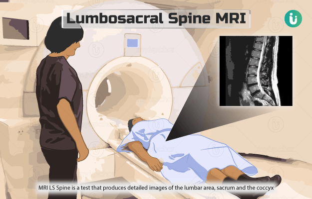
A magnetic resonance imaging (MRI) scan is an imaging test that generates three-dimensional pictures of the body structures and organs using radio waves and strong magnetic fields. This test helps detect and diagnose diseases and monitors ongoing treatment. Some of the machines used to conduct an MRI are spacious while some are like a narrow tunnel. The test can last anywhere from 30 minutes to 2 hours depending on the suspected disease or condition.

The magnet in an MRI machine is measured in the unit Tesla (T). The commonly used magnet in an MRI machine is 1.5T; however, the latest advancement, the 3T MRI machine, has a magnet strength of 3T, which has a stronger signal. This leads to improved image quality with a decrease in test time.

Normally, you will have to lie down on an examination table, which will then slide into a tunnel-like machine that contains the magnets. However, since the MRI procedure takes a long time, it can cause discomfort to the patients (especially those who are claustrophobic). Advancement in technology has greatly helped to minimise this. 

In a reclining MRI, the patient can rest in a reclining chair with only the part of the body that is to be studied inside the equipment. No other body part is in the machine. During the scan, the patient may either choose to read a book or listen to music. Since the patients are not inside the MRI machine, they do not need to worry about claustrophobia.

Additionally, some MRI machines with an advanced design allow imaging of different parts of the body such as bones, cartilages and joints while standing, squatting or in other weight-bearing positions. Such a machine is known as an upright open MRI and is beneficial in understanding the functioning of the joint. When there is injury or damage to the joints, the joint can be scanned in a position relevant to normal joint function as well as through the range of motion of the joint.

  1. Why is Magnetic Resonance Imaging test performed?
  2. How do you prepare for a Magnetic Resonance Imaging Test?
  3. How is a Magnetic Resonance Imaging Test performed?
  4. What do the test results of an MRI test mean?

MRI test, when used with other imaging techniques, can help diagnose various health conditions. In some MRIs, contrast dyes (often containing gadolinium, an element) are used to obtain more information about blood vessels in a particular area. 

  • MRI is suitable for imaging of soft tissues and non-bony areas of the body. It generates better images of tendons, muscles, brain, ligaments, nerves, and spinal cord as compared to computed tomography (CT) scan and X-rays.
  • This imaging helps differentiate between the grey and white matters of the brain and detects tumours and aneurysms (an outward bulge) in the brain.
  • An MRI scan usually gives more accurate information about shoulder and knee injuries and conditions of the brain and spinal cord, e.g., venous malformations (abnormalities in the veins), hydrocephalus (presence of fluid in the brain), haemorrhage, and abscesses.
  • Functional magnetic resonance imaging (fMRI) is a specialised MRI technique that helps detects the active parts of brain while the person is performing tasks that activate certain areas in brain.

As MRI scans do not use damaging radiations, they are suitable for diagnosing diseases and injuries that require repeated imaging.

Upright open MRI is also used to understand joint motion and alignment, especially in disease conditions such as femoroacetabular impingement (a condition where there is extra bone growth along the bones of the hip joint, giving it an irregular shape) and osteoarthritis.

A 3T MRI has the following benefits over a 1.5T MRI:

  • Better image quality in different organs such as the spine, heart, breast, blood vessels and neuromuscular system.
  • Improved imaging of brain lesions that occur in multiple sclerosis
  • Reduced scan time
  • Can help in visualising the small organs in paediatric patients.
  • A 3T MRI is more sensitive to gadolinium dye compared to 1.5T MRI, thus requiring a lesser dose of the dye or providing better quality images in the same dose.
myUpchar doctors after many years of research have created myUpchar Ayurveda Urjas Capsule by using 100% original and pure herbs of Ayurveda. This ayurvedic medicine has been recommended by our doctors to lakhs of people for sex problems with good results.
Long Time Capsule
₹712  ₹799  10% OFF
BUY NOW

To prepare for an MRI, you might be asked not to drink or eat anything for 4 to 6 hours before the test. You will be asked to wear a hospital gown and put away your eyeglasses, pens, hairpins, credit cards, dental objects, hearing aids and jewellery in a locker. It is advisable to keep all your valuable items at home before coming for the test.

While taking an appointment for an MRI scan, remember to inform the physician if you have any of the following conditions:

  • Permanent tattoos and eyeliner or body piercings
  • Metal implants such as metal rods, screws and plates, and surgical staples
  • Pregnancy
  • A medical patch on your body
  • Difficulty in lying down for about 30 to 60 minutes
  • Previous bullet wound
  • Recent placement of any artificial joints

Following steps are generally followed in an MRI test:

  • If advised by the physician, a contrast dye is injected through an intravenous drip placed in the arm or hand.
  • You will be asked to lie down on a table, which then slides into the spherical opening of the MRI scanning machine. Pillows or straps may be used to secure your position and avoid any movements during the scan.
  • The technician conducting the MRI scan will be in a different room with scanner controls. The technician will be able to monitor you through a window connecting the rooms.
  • You can communicate with the technician by pressing a call button. In case of any difficulty during the procedure, communication is also possible from inside the scanner.
  • You will be given a headset with or without music or earplugs to block the noise made by the scanner. You might hear a click during the scan once the radio wave pulses and magnetic field release from the scanner.
  • If required, the technician might instruct you to hold your breath for a few seconds. Do not resume breathing unless you are asked to.
  • To achieve accurate results from the scan, it is essential to stay still during the scan. The slightest movement can create false images and affect the quality of the scan.

In an upright open MRI, you may need to sit, stand or lie down. You may have to change your postures during the scan. Similarly, for a reclining MRI you will be sitting in a reclining chair while the machine scans the concerned part of your body. Hold as still as you can during the scan. It takes two to four minutes while standing and five to 10 minutes when you are sitting or lying down.

An MRI does not produce any side effects from the radio waves and magnetic fields. Allergic reactions to the contrast dye occur in extremely rare cases. Magnetic fields generated while scanning can affect the working of implanted devices such as pacemakers and cochlear implants; therefore, it is essential to inform your physician of such implants before an MRI scan. Gadolinium, the most commonly used dye in MRI scans, is harmful to people with kidney problems. So, you should inform the technician if you have kidney problems.

Normal results:

A normal MRI scan represents that the body part being examined in the scan is not suffering from any health condition.

Abnormal results:

Results of an MRI scan are interpreted on the basis of signals received from the body part examined during the scan. For instance, cancer tissue cells provide different signals as compared to healthy tissue cells. Consulting your health care provider with the results will help resolve any queries and concerns about the results.

Disclaimer: This information is purely from an educational point of view and is in no way a substitute for medical advice by a qualified doctor.  All results must be clinically correlated with the patient’s complaints to make a complete and accurate diagnosis.

References

  1. Magnetic resonance imaging (MRI) test; John Hopkins Medicine, John Hopkin's University; c2019
  2. Carpenter JP, Litt H, Gowda M. Magnetic resonance imaging and arteriography. In: Sidawy AN, Perler BA, eds.Rutherford's Vascular Surgery and Endovascular Therapy. 9th ed. Philadelphia, PA: Elsevier; 2019:chap 28.
  3. Wilkinson ID, Graves MJ. Magnetic resonance imaging. In: Adam A, Dixon AK, Gillard JH, Schaefer-Prokop CM, eds. Grainger & Allison's Diagnostic Radiology. 6th ed. Philadelphia, PA: Elsevier; 2015:chap 5.
  4. National Institute of Biomedical Imaging and Bio engineering [internet]: US Department of Health and Human Services; Magnetic Resonance Imaging (MRI)
  5. Medline plus [internet]: US National Library of Medicine; MRI scans
Read on app
cross
Ask your health query from live doctors now!