What is a Follicular Study Ultrasound?
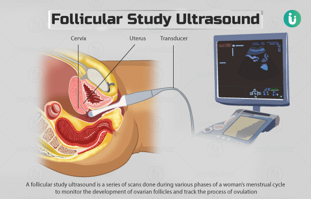
A follicular study ultrasound, also known as follicular tracking or follicular monitoring, is a series of scans done during various phases of a woman’s menstrual cycle to monitor the development of ovarian follicles and track the process of ovulation. This test also checks the endometrial changes in response to maturation of the eggs. It is mainly used to help couples to time intercourse - to the exact time of ovulation - and optimise the chances of having a baby. Endometrium is the inner wall of the uterus.
Ovaries—a pair of female reproductive organs—contain thousands of tiny fluid-filled sacs called follicles in which eggs grow. During the start of a woman’s menstrual cycle, several of these follicles begin to develop. However, by midcycle, only one ‘dominant’ follicle will have developed enough to break open and release a mature egg. The stage of the menstrual cycle when the ovary releases a mature egg is called ovulation.
The egg is picked up by the fallopian tube and then waits in the fallopian tube for possible fertilisation (fusion with a sperm to form a new life). During the whole process, the endometrium also changes in thickness. Around the time of ovulation, the endometrial lining starts to thicken and gets more blood supply. This is to ensure implantation in case fertilisation occurs.
Follicular study is performed by a transvaginal ultrasound. In this method, a sterile (clean) probe or transducer covered with a probe cover (usually a condom) is inserted into the vagina. The probe emits sound waves and captures the waves that are reflected off the internal organs. The ultrasound machine uses these reflected waves to create clear images of the uterus and ovaries on a screen, with particular attention given to the follicles present in ovaries.


