What is a Computed Tomography (CT) Scan of Paranasal Sinuses (PNS) (Axial and Coronal)? 

A PNS CT scan is an imaging test that helps check your paranasal sinuses, which are air-filled hollow spaces in the bones around your nasal cavity.

A CT scan uses x-ray radiation and a computer to generate slices (cross-sectional images) of your internal organs, bones, blood vessels and soft tissues. The images of the PNS can be viewed either in axial or coronal position. 

  • Coronal view:

    • For a coronal view, the individual is asked to lie face down on the CT scanning table with their hard palate (the hard portion in the roof of the mouth) perpendicular to the gantry of the scanning table. Gantry is the circular frame into which the person slides during the CT scan.
    • A coronal view will give images of the sinuses from front to the back.
  • Axial view:
    • In the axial view, the person is asked to keep their hard palate perpendicular to the scanning table.
    • It gives a view of the sinus cavities from top to bottom.
    • If a person is not able to lie face down (in prone position) for the coronal scan, a computer-generated reconstructed coronal view can be obtained from axial sections.

Although this test is performed without using a contrast dye, in some situations, the doctor may order a CT scan with contrast dye to get detailed images of structures inside the body.

  1. Who cannot have a PNS CT Scan (Axial/coronal)?
  2. Why is a PNS CT Scan (Axial/coronal) done?
  3. How should I prepare for a PNS CT Scan (Axial/coronal)?
  4. How is a PNS CT Scan (Axial/coronal) done?
  5. How will a PNS CT Scan (Axial/coronal) feel like?
  6. What do the results of a PNS CT Scan (Axial/coronal) mean?
  7. What are the risks and benefits of a PNS CT Scan (Axial/coronal)?
  8. What happens after a PNS CT Scan (Axial/coronal)?
  9. Contrast vs Non-contrast CT Scan PNS
  10. What are the other tests that can be done with a PNS CT Scan (Axial/coronal)?

CT scan is avoided in pregnant women as the radiation can harm the unborn baby.

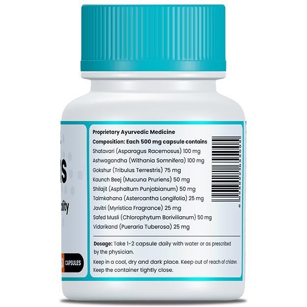
myUpchar doctors after many years of research have created myUpchar Ayurveda Urjas Capsule by using 100% original and pure herbs of Ayurveda. This ayurvedic medicine has been recommended by our doctors to lakhs of people for sex problems with good results.
Long Time Capsule
₹712  ₹799  10% OFF
BUY NOW

This test is performed to:

  • Help with planning or checking the paranasal sinus after a sinus surgery 
  • Check for tumours, infections, polyps or injury in the sinus cavity
  • Detect birth defects related to the sinuses

Your healthcare practitioner may order this test if you show some of the following symptoms related to the abovementioned conditions:

The following preparations are needed before a CT scan PNS axial/coronal:

  • You need to wear loose and comfortable clothes for the test. 
  • A technologist may provide you with a hospital gown right before the test. 
  • You will be asked to remove jewellery, hairpin, eyeglasses, removable dentures and hearing aids.
  • Fasting is required if the contrast dye is to be used. 
  • Tell your healthcare provider if you have the following conditions:

The following steps are performed for a PNS CT scan:

  • The healthcare provider may ask you to lie down either on your back or in a prone position. 
  • If a contrast dye is required, they will inject it through an intravenous line in a vein in your arm. 
  • During the scan, the table will slide into the tunnel-like circular path. 
  • You need to hold your breath when the images are captured. 
  • After the test, the technologist will assist you off the table.

The entire procedure takes about 10 minutes.

myUpchar doctors after many years of research have created myUpchar Ayurveda Kesh Art Hair Oil by using 100% original and pure herbs of Ayurveda. This Ayurvedic medicine has been recommended by our doctors to more than 1 lakh people for multiple hair problems (hair fall, gray hair, and dandruff) with good results.
Bhringraj Hair Oil
₹546  ₹850  35% OFF
BUY NOW

CT scan for PNS is a painless test. However, you may feel uncomfortable lying down still for the test. When the dye is injected, you may feel:

  • Metallic taste in the mouth 
  • Burning sensation
  • Warm flushing of the body

The results in the CT scan for PNS may indicate the following conditions:

 The benefits of doing a CT scan are as follows:

  • It is a painless and non-invasive method. 
  • It can be helpful for people with implanted devices, unlike MRI
  • There are no immediate side effects of x-ray radiation.

The risks of the CT scan are as follows:

  • In rare cases, people are allergic to the contrast dye.

myUpchar doctors after many years of research have created myUpchar Ayurveda Urjas Energy & Power Capsule by using 100% original and pure herbs of Ayurveda. This Ayurvedic medicine has been recommended by our doctors to lakhs of people for problems like physical and sexual weakness and fatigue, with good results.
Power Capsule For Men
₹495  ₹799  38% OFF
BUY NOW

You will be able to do your normal activities after the test.

A non-contrast CT scan PNS is generally done, except in cases like complicated acute sinusitis (e.g., periorbital cellulitis or abscess) where a CT scan with contrast is ordered.

If contrast is required, you may need to fast for the test. When the dye is injected, you may feel warm flushing and may have a metallic taste in your mouth. Rarely, some people are allergic to the contrast dye.

Depending on the condition, the doctor may order other tests that may include the following:

Disclaimer: All results must be clinically correlated with the patient’s complaints to make a complete and accurate diagnosis. The above information is provided from a purely educational point of view and is in no way a substitute for medical advice by a qualified doctor.

References

  1. Minni A, Messineo D, Attanasio G, Pianura E, D'Ambrosio F. 3D cone beam (CBCT) in evaluation of frontal recess: findings in youth population. Eur Rev Med Pharmacol Sci. 2012 Jul. 16(7): 912-8. PMID: 22953640.
  2. Aksoy EA, Ozden SU, Karaarslan E, et al. Reliability of high-pitch ultra-low-dose paranasal sinus computed tomography for evaluating paranasal sinus anatomy and sinus disease. J Craniofac Surg. 2014 Sep. 25(5): 1801-4. PMID: 25203576.
  3. Tomovic S, Esmaeili A, Chan NJ, Choudhry OJ, Shukla PA, Liu JK, et al. High-resolution computed tomography analysis of the prevalence of Onodi cells. Laryngoscope. 2012 Jul. 122(7):1470-3. PMID: 22685058.
  4. Pirimoglu B, Sade R, Sakat MS, Ogul H, Levent A, Kantarci M. Low-Dose Noncontrast Examination of the Paranasal Sinuses Using Volumetric Computed Tomography. J Comput Assist Tomogr. 2018 May/Jun. 42 (3): 482-6. PMID: 29287024.
  5. Al Abduwani J, ZilinSkiene L, Colley S, Ahmed S. Cone beam CT paranasal sinuses versus standard multidetector and low dose multidetector CT studies. Am J Otolaryngol. 2016 Jan-Feb. 37(1): 59-64. PMID: 26700263.
  6. Chakravarti A, Naglot S, Dhawan R. Outcome of endoscopic sinus surgery in patients with symptomatic chronic rhinosinusitis with minimal changes on computerised tomography. Indian J Otolaryngol Head Neck Surg. 2011 Oct. 63(4):359-63. PMID: 23024942.
  7. Cebula M, Danielak-Nowak M, Modlinska S. Impact of Window Computed Tomography (CT) Parameters on Measurement of Inflammatory Changes in Paranasal Sinuses. Pol J Radiol. 2017. 82: 567-70. PMID: 29662587.
  8. Yousem DM. Imaging of sinonasal inflammatory disease. Radiology. 1993 Aug. 188(2): 303-14. PMID: 8327669.
  9. Radiological Society of North America (RSNA) [internet]. Oak Brook. Illinois. USA; Computed Tomography (CT) - Sinuses
  10. National Cancer Institute. [Internet]. National Institute of Health. U.S. Department of Health & Human Services; Anatomical Terminology.
  11. Nichols JR, Puskarich MA. Abdominal trauma. In: Walls RM, Hockberger RS, Gausche-Hill M, eds. Rosen's Emergency Medicine: Concepts and Clinical Practice. 9th ed. Philadelphia, PA: Elsevier; 2018:chap 39.
  12. Herring W. Recognizing the normal abdomen and pelvis on computed tomography. In: Herring W, ed. Learning Radiology: Recognizing the Basics. 4th ed. Philadelphia, PA: Elsevier; 2020:chap14
  13. Chernecky CC, Berger BJ. Computed tomography of the body (spiral [helical], electron beam [EBCT, ultrafast], high resolution [HRCT], 64-slice multidetector [MDCT])-diagnostic. In: Chernecky CC, Berger BJ, eds. Laboratory Tests and Diagnostic Procedures. 6th ed. St Louis, MO: Elsevier Saunde
  14. O'Handley JG, Tobin EJ, Shah AR. Otorhinolaryngology. In: Rakel RE, Rakel DP, eds. Textbook of Family Medicine. 9th ed. Philadelphia, PA: Elsevier Saunders; 2016:chap 18
  15. Pappas DE, Hendley JO. Sinusitis. In: Kliegman RM, Stanton BF, St. Geme JW, Schor NF, eds. Nelson Textbook of Pediatrics. 20th ed. Philadelphia, PA: Elsevier; 2016:chap 380
  16. Murr AH. Approach to the patient with nose, sinus, and ear disorders. In: Goldman L, Schafer AI, eds. Goldman-Cecil Medicine. 25th ed. Philadelphia, PA: Elsevier Saunders; 2016:chap 426
  17. Rosenfeld RM, Piccirillo JF, Chandrasekhar SS, Brook I, Ashok Kumar K, Kramper M, Orlandi RR, Palmer JN, Patel ZM, Peters A, Walsh SA, Corrigan MD. Clinical practice guideline (update): adult sinusitis. Otolaryngol Head Neck Surg. 2015 Apr;152(2 Suppl):S1-S39. PMID: 25832968
  18. DeMuri GP, Wald ER. Sinusitis. In: Bennett JE, Dolin R, Blaser MJ, eds. Mandell, Douglas, and Bennett's Principles and Practice of Infectious Diseases, Updated Edition. 8th ed. Philadelphia, PA: Elsevier Saunders; 2015:chap 63
  19. Soler ZM, Smith TL. Results of medical and surgical treatment of chronic rhinosinusitis with and without nasal polyps. In: Flint PW, Haughey BH, Lund V, et al, eds. Cummings Otolaryngology: Head & Neck Surgery. 6th ed. Philadelphia, PA: Elsevier Saunders; 2015:chap 44
  20. Bachert C, Calus L, Gevaert P. Rhinosinusitis and nasal polyps. In: Adkinson NF, Bochner BS, Burks AW, et al, eds. Middleton's Allergy: Principles and Practice. 8th ed. Philadelphia, PA: Elsevier Saunders; 2014:chap 43
  21. Rosenfeld RM, Piccirillo JF, Chandrasekhar SS, Brook I, Kumar KA, Kramper M, Orlandi RR, Palmer JN, Patel ZM, Peters A, Walsh SA, Corrigan MD. Clinical practice guideline (update): Adult Sinusitis Executive Summary. Otolaryngol Head Neck Surg. 2015 Apr;152(4): 598-609. PMID: 25833927
  22. Okuyemi KS and Tsue TT. Radiologic Imaging in the Management of Sinusitis. Am Fam Physician. 2002 Nov 15;66(10): 1882-1887. PMID: 12469962.
Read on app
cross
Ask your health query from live doctors now!