Skin ulcer

Dr. Srishti GuptaMBBS

June 08, 2021

June 08, 2021

Skin ulcer
Skin ulcer

An ulcer can be defined as a break in the skin or mucous membrane with loss of surface tissue, disintegration and necrosis (or death) of epithelial tissue and often exudation of pus. Ulcers can occur in any part of the body that has an epithelial lining. In order to understand the different appearance, types and healing of ulcers it is important to know that all ulcers, irrespective of their location, are composed of four parts – the base, the floor, the edge and the margin. The base of the ulcer is the tissue upon which it lies and it can be soft tissue or bone. The floor of the ulcer differs from the base, it overlies the base and is the deepened central portion containing the dead cell debris, pus and granulation tissue. While the margins of an ulcer refer to its boundaries, the edge is the portion that connects the depressed floor to the margin. Characteristics of the edge of the ulcer are of paramount importance as they are its distinguishing feature. One type of ulcer can be differentiated from another on the basis of its edges alone.

Skin ulcers simply refer to ulcers that occur on the skin. Unlike organic ulcers like peptic ulcers, ulcers of the skin are noticeable to the naked eye. Sometimes, skin ulcers are confused with skin rashes or other lesions. A skin rash is an area of the skin that has changed in texture or colour and may look inflamed or irritated. The skin may be red, warm, scaly, bumpy, dry, itchy, swollen, painful, or may have cracks and blisters. A rash can be widespread and occur in multiple distant areas of the body at the same time, unlike an ulcer, which is usually confined to one region. Skin rashes are not associated with a break in the epithelium, as with ulcers, of the skin and can have many other characteristic varieties based on the type of skin lesion – macules, papules, plaques, vesicles, pustules and bullae to name a few. Skin rashes can, however, lead to skin ulcers or be accompanied by them, in certain skin conditions. Even though the term skin ulcers implies a dermatological disease, ulceration of skin can occur secondary to other systemic diseases (like autoimmune diseases) and even invasive cancer. Skin ulcers can be categorised by their cause and location. Some are primary problems and some are manifestations of another, sometimes more serious, underlying disease.

Edges of skin ulcers

Based on the edge of the ulcer, it can be typified. This information is important for examining, identifying and correctly diagnosing the ulcer:

  • Sloping edge ulcer: The edge between the margin and the floor of the ulcer may be at a sloping incline; this usually implies the ulcer is healing. Besides healing ulcers, this is also seen in venous ulcers and ulcers resulting from trauma
  • Punched out edge ulcer: This type of ulcer tends to be quite deep and almost appears as if it has been punched out. This crater-like appearance of an ulcer can be found in ulcers that do not heal (non-healing ulcers) and is also seen with gummatous ulcers of syphilis and trophic ulcers.
  • Undermined edge ulcer: In some ulcers, the disease tends to destroy the subcutaneous tissue underlying the skin faster than it destroys the skin. The remaining unhealthy skin margins overhang over the edge of the ulcer, giving it the diagnostic undermined edge appearance. Skin ulcers seen in tuberculosis are typical examples of such ulcers. 
  • Raised edge ulcer: In this variety of ulcers, the edge seems to be raised like a bump where it meets the margin of the ulcer. This is a very characteristic sign of basal cell carcinoma, a type of ulcerative skin cancer that affects the head, especially the face and neck.
  • Everted edge ulcer: Some ulcers have edges that roll out or become everted at the margins. Ulcers with rolled out or everted edges at the margins are a hallmark of squamous cell carcinoma, another more aggressive type of skin cancer. 
Face Serum
₹349  ₹599  41% OFF
BUY NOW

Signs and symptoms of skin ulcers

Usually, skin ulcers are associated with other symptoms specific to their underlying disease that could include, but are not limited to:

  • Bulging lower limb veins
  • Loss of sensation or tingling in the feet
  • High blood pressure

Skin ulcers differ in presentation and appearance depending on the type but some features are common to most. These are:

Types and causes of skin ulcers

Ulcers can occur in any part of the body lined by a mucosal membrane or an epithelial lining. Therefore, skin ulcers are very common. Skin ulcers can be classified in various ways, including on the basis of location, type of disease and potential to heal.

  • Types of ulcers on the basis of healing capability:
    • Spreading: The ulcer-causing underlying disease process is active and ulceration activity is ongoing in such ulcers. The skin surrounding the ulcer shows signs of inflammation – redness, warmth, tenderness and swelling.
    • Healing: The ulcerative process has terminated and these ulcers have begun to heal. The edges tend to be sloping.
    • Callus: Ulcers that do not heal, and show no tendency to, have pink granulation tissue in their floor and often have punched out edges.
  • Types of ulcers on the basis of disease or causes:
    • Venous ulcers: Venous ulcers typically occur on the legs, especially below the knees and along the inner medial surface. They are quite common near the ankles, especially the medial malleolus, which is the bony projection on the inner medial surface of the ankle. The function of the veins of the legs is to pump the blood back towards the heart against the force of gravity. Sometimes, the valves that prevent the backflow of blood in these veins fail and the pressure of blood in the calves and legs increase. The skin of the legs becomes fragile under this added pressure and ulcers can develop, especially at bony points in the leg. These ulcers are usually shallow and do not penetrate very deep. They are not associated with pain unless they become infected. Following are some risk factors for developing venous ulcers: 
    • Arterial ulcers: Unlike venous ulcers, arterial ulcers are usually very painful and penetrate deep into the fascia. Arterial ulcers are also called ischaemic ulcers and occur due to problems in the arterial blood circulation, most commonly of the legs, depriving the tissues of necessary oxygen and leaving them vulnerable to damage. Arterial ulcers can occur in young and old patients. Causes and risk factors of arterial ulcers include: 
    • Trophic or neuropathic ulcers: Sometimes certain diseases cause neuropathy and lead to the patient losing sensation (touch, temperature and pain) in parts of the body (most commonly of the feet). As the patient can not feel it, repeated trauma occurs and goes unnoticed, especially in the bony joints and prominences of the foot. First callosities are formed over bony points like ankles and toes, then they become infected and pus forms. This type of perforating ulceration burrows deep through the tissues, sometimes even reaching and infecting the underlying bone. Examples of causes of neuropathic ulcers include: 
      • Uncontrolled diabetes mellitus
      • Leprosy
      • Alcoholic neuropathy
      • Nerve injuries
      • Transverse myelitis
    • Pressure ulcers: Commonly called bed sores, and also known as decubitus ulcers, these develop due to prolonged pressure over bony prominences, like that in the buttocks and back. These commonly occur in patients battling ailments who are bedridden for long periods of time. Therefore, it is necessary to prevent them by moving the patient periodically.
    • Tropical ulcers: Also called phagedenic ulcer, this ulcer may develop in the feet of people in tropical climates who do not wear footwear, live in poor sanitation, are exposed to factors like insect bites and suffer from intestinal parasites. The feet appear gnawed on.
    • Cancer-associated ulcers: While some cancers of other organs can invade and extend into the skin at more advanced stages (like breast cancer), cancers of the skin can also sometimes present as non healing ulcers. Causes of skin-cancer-related ulcers include: 
      • Basal cell carcinoma: Also known as rodent ulcer, is the most common type of skin cancer. It appears as pearly pink ulcers on the head and neck.
      • Squamous cell carcinoma of the skin
      • Melanoma
    • Uncommon ulcers: Some less common skin ulcers are also possible and are caused by specific circumstances:
      • Martorell's ulcer: Also known as Martorell’s hypertensive arteriosclerotic ischemic ulcer, it is a very painful ulcer of the legs that occurs in patients with long-standing poorly controlled, or uncontrolled, high blood pressure.
      • Meleney's ulcer: This is a type of ulcer with undermining edges that affects the skin and subcutaneous tissue at the site of operation. It arises due to a postoperative infection at a surgical site by multiple organisms.
      • Bazin’s ulcer: Young women suffering from tuberculosis can have nodular eruptions in the back of their calves which become ulcerated. This type of panniculitis (inflammation of the fat underlying the skin) is known as erythema induratum or Bazin’s ulcers. It is different from erythema nodosum which is a nodular eruption that occurs on the shins, and not the calves, of young women with tuberculosis.
      • Marjolin ulcer: A Marjolein ulcer is a rare and aggressive type of skin cancer that grows from burns, scars or keloids. It appears like a non-healing sore in scar tissue.

Diagnosis of skin ulcers

The doctor begins by taking a thorough medical history that focuses on when the ulcers appeared, how they progressed and associated symptoms (like pain or itching). After noting the present complaints, the doctor will also take a history of preexisting health conditions like diabetes mellitus, varicose veins or documented peripheral arterial disease. Personal history (like habit of smoking) is important to disclose to the healthcare team to reach an accurate diagnosis. Following a detailed medical history, the doctor will begin a comprehensive physical examination of the patient. Starting with general physical examination, other systems of the patient will be evaluated. The ulcer is evaluated by palpation with gloved hands. It is important for the doctor to identify the type of ulcer by noting the base, floor, edges and margins of the ulcer. This process may not hurt but may cause minor discomfort to the patient. Some clinical tests for varicose veins using tourniquets are also usually performed. Neurological testing is essential, especially in a known diabetic patient, as neuropathy (marked by lack of sensation in the feet) is the usual culprit in diabetic ulcer foot disease. After fully evaluating the patient in the clinic, some blood tests, radiological imaging studies and, at times, skin biopsies are ordered to arrive at a definitive diagnosis.

Skin Infection Tablet
₹719  ₹799  10% OFF
BUY NOW

Tests for skin ulcers

Blood tests and some radiological imaging studies are generally useful in correctly diagnosing the cause of skin ulcers.

  • Blood tests:
  • Radiological imaging: 
    • X-rays: Affected area of the ulcer is visualised to see if there is an extension into the bone.
    • Doppler ultrasound: A handheld device that uses ultrasound waves is used to detect the blood flow in arteries of the lower limb. Ankle-brachial pulse index (ABPI) is calculated; if it is less than 1, arterial flow is considered compromised. 
  • Skin biopsy: In cases of suspected skin cancer, skin biopsy, usually a punch biopsy, is taken by the doctor. Histological studies are done on it to detect cancer cells. 

Management of skin ulcers

Treatment of the ulcer focuses on the treatment of the underlying cause. Usually, after the disease is managed, the ulcers resolve. However, sometimes ulcers need special care to overcome the existing damage.

  • Dressing: The type of dressing to be done depends on the type of skin ulcer. Dead tissue, dirt, debris or pus is usually removed while dressing any type of ulcer. Some ulcers need to be cleaned at each dressing and warm water or normal saline (NS) is typically used. Some key points about dressing skin ulcers include: 
    • Non-sticky dressings are used for painful ulcers
    • Ulcers at added risk of skin infection are dressed with antiseptic dressing or ointment 
    • Ulcers that bleed a lot can benefit from absorptive alginate dressings
    • Venous leg ulcers due to varicose veins are treated with compression bandages in addition to dressings to restore venous blood flow
  • Medicines: Painkillers are advisable to help with the pain. Antibiotics may be prescribed if the ulcer is infected. Steroid creams are used to reduce itching.

Complications associated with skin ulcers

Untreated or extensive skin ulcers pose the risk of causing other complications which include:

  • Infections: The weakened skin of the ulcer can easily become infected.
  • Cellulitis: Infected ulcers can penetrate deeper and spread the infection to subcutaneous tissues.
  • Osteomyelitis: Aggressive infected skin ulcers, like trophic ulcers of diabetic ulcer foot disease, can even burrow all the way to the underlying bones to cause bone infection.
  • Sepsis: Lastly, the infection may spread into the bloodstream to cause sepsis and even septic shock in some cases.
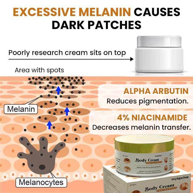
Body Brightening Cream
₹349  ₹649  46% OFF
BUY NOW

Prognosis of skin ulcers

The outlook of skin ulcers depends on their underlying cause and the overall health of the patient. Not only does the severity and type of the underlying disease determine prognosis, but the extent of ulceration and healing potential controls the overall cosmetic result. Some ulcers may heal completely without leaving a scar, whereas others may never fully heal.


Medicines for Skin ulcer

Medicines listed below are available for Skin ulcer. Please note that you should not take any medicines without doctor consultation. Taking any medicine without doctor's consultation can cause serious problems.

Medicine Name

Price

₹210.0

₹90.25

Showing 1 to 0 of 2 entries