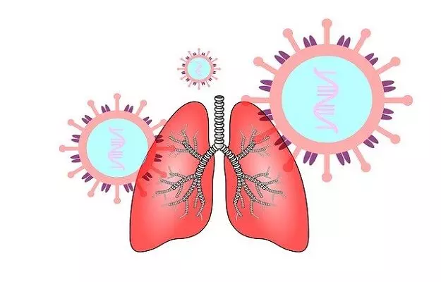
COVID-19 is an acute respiratory disease that mainly affects the airways and lungs. The virus binds to specific receptors called the ACE2 receptors in the lungs. These receptors are otherwise needed to maintain blood pressure and fluid balance in the body - but the SARS-CoV-2 virus, the causative agent of COVID-19, uses them to enter healthy lung cells and cause infection. (Read more: What are ACE2 receptors and what do they have to do with COVID-19)
The disease presents as a variety of symptoms including cough, fever and shortness of breath. (Read more: Mild vs Severe COVID-19)
However, symptoms are not the only thing used to diagnose this disease. COVID-19 is mostly diagnosed through the RT-PCR test but doctors also look for symptoms of the disease through radiological testing like CT scans in areas where there is widespread infection. The scan looks for specific changes in the lungs that are seen in COVID-19 patients.
A CT scan of the chest is preferred over other radiographs of the chest (like chest X-ray) as it is more sensitive and is helpful in identifying the early changes in COVID-19. Here is what the CT scans of COVID-19 patients look like and how the disease affects the lungs:
CT scans of lungs of COVID-19 patients
Experts suggest that chest CT scans of COVID-19 patients don’t have many unique signs: most of the signs that are seen in COVID-19 are also seen in other lung diseases.
That said, there are certain things that can be seen in a lung CT scan of COVID-19 patients that can point to possible infection - especially if the patient is at risk of exposure to the virus. People living in badly affected areas or countries, caregivers to COVID-19 patients at home and medical professionals on the frontlines of the war against COVID-19 are all considered to have higher risk of infection.
Most commonly a COVID-19 patient shows bilateral nodular lesions with surrounding ground-glass opacities in CT scan. Also, consolidations are seen. Here's what this means:
- Ground glass opacities: In radiology, ground-glass opacities are hazy areas in lungs that are clear enough to still be able to see the underlying bronchial structures. Ground glass opacities may point to a number of conditions including inflammation, collapse of alveoli (air sacs present in lungs), oedema, and fibrosis. (Read more: Inflammation and COVID-19)
- Consolidations: Consolidations are much denser than ground-glass opacities and make it impossible to see the structures under them. Consolidations usually occur due to the presence of liquid or solid in the lungs or airways.
A healthy lung would look completely black in a CT scan.
A CT scan is an imaging technique which uses X-ray radiation to produce images on the internal structures of the body. The CT scan machine has a circular scanner that rotates around the person’s body and sends X-ray radiations. These radiations create slice images (tomographic slices) which are then combined (by an attached computer) to make a 3D image of the scanned area.
In a recent study published in the Journal of Thoracic Oncology, a group of health practitioners from China mentioned two case studies in which patients who had recently had surgery for lung cancer showed oedema, fibrosis, and inflammation. Both the patients were later found to have COVID-19.
Read more: Who can get tested for COVID-19


