Summary
Valve replacement surgery is used to replace a diseased or damaged valve in the heart. It can be either open-heart or minimally invasive surgery. Valves are flaps of tissue that prevent the backflow of blood in heart. Any problem in a heart valve could cause health conditions. Valve replacement surgery helps to relieve symptoms of a damaged valve and prolongs your life.
Please click on this link to know a better heart disease diet plan.
Before a valve replacement surgery, healthcare practitioners perform certain tests to check if you are fit for the surgery. The operation will require general anaesthesia. After the surgery, you will be asked to keep the area of the incision clean and dry and to take special precautions like avoiding lifting heavy objects or moving your upper body too much. In addition, a proper diet, routine exercise and regular medication will help you in speedy recovery after surgery.
(Read More - First Aid for Heart Attack)
- What is heart valve replacement surgery?
- Why is heart valve replacement surgery recommended?
- Who can and cannot get a heart valve replacement surgery?
- What preparations are needed before a heart valve replacement surgery?
- How is a heart valve replacement surgery done?
- How to care for yourself after a heart valve replacement surgery?
- What are the possible complications/risks of a heart valve replacement surgery?
- When to follow up with your doctor after a valve replacement surgery?
What is heart valve replacement surgery?
The heart is a pump that is divided into four chambers (two atria, the upper chambers, and two ventricles, the lower chambers).
These chambers are connected with valves to keep the blood flowing in one direction. The valves open and close on each heartbeat and prevent backflow of the blood. Valves are also located at the large blood vessel (aorta) that controls the flow of blood out of the heart to the rest of the body.
There are four such valves present in the heart:
- Mitral valve - located between left atrium and left ventricle
- Tricuspid valve - located between right atrium and right ventricle
- Pulmonary valve - located between right ventricle and pulmonary artery
- Aortic valve - located between left ventricle and aorta
Damage to any of the heart valves can affect the blood flow through the heart. If a valve does not open completely, it will obstruct the flow of blood (valve stenosis). If the valve does not close fully, it can cause blood to leak backwards (valve regurgitation).
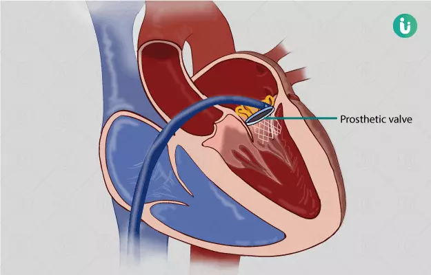
Valve replacement surgery is usually done on the aortic or mitral valve. The surgery is performed to replace a severely damaged or diseased valve when one or more valves are affected. It is also helpful in treating valve diseases that can be fatal. There are two types of valves used for replacement:
- Mechanical valves: These valves are made of plastic, carbon, or metal. Mechanical valves last longer than biological valves and are durable. However, blood can stick to the valves and form blood clots; therefore, blood-thinning medications are given to individuals with mechanical valves to prevent blood clots.
- Biological valves: These valves are made from animal tissue or human tissue from a donated heart. Sometimes, a person’s own tissue can be taken to replace the valve. People with biological valves do not need blood-thinning medications. Biological valves are not tougher than mechanical valves and need to be replaced every 10 years.
Heart valve replacement surgery can be performed in two ways:
- Open-heart surgery: This is a traditional method in which a large incision (cut) is made in the chest, and the heart is stopped for some time to replace the valve.
- Minimally invasive surgery: This involves smaller incisions to replace the heart valve. There is less pain after minimally invasive surgery, and the hospital stay and recovery time are also shorter compared to open-heart surgery.
(Read More - Peripheral artery disease)
Why is heart valve replacement surgery recommended?
Your doctor may recommend a valve replacement surgery if you have a damaged or diseased valve and show the following symptoms:
- Palpitations
- Chest pain
- Difficulty in breathing
- Dizziness
- Weight gain due to fluid retention
- Swelling in the feet, ankle, or abdomen
(Read More - Myocarditis treatment)
Who can and cannot get a heart valve replacement surgery?
Valve replacement surgery is recommended in people with the following conditions:
- Aortic insufficiency
- Congenital (present from birth) heart valve disease
- Aortic valve stiffness (stenosis)
- Mitral valve stenosis
- Congenital heart valve disease
- Mitral regurgitation - acute
- Mitral regurgitation - chronic
- Pulmonary valve stenosis
- Tricuspid regurgitation
- Tricuspid valve stenosis
Traditionally, open-heart surgery is performed for valve replacement. However, old individuals with co-existing conditions such as lung or kidney disease, peripheral vascular disease (a blood circulation disorder) or diabetes, are at increased risk of complications from the surgery. In such individuals, minimally invasive heart valve replacement surgery may be performed.
Minimally invasive surgery has its own limitations as it cannot be done in people with the following conditions:
- Severe valve damage
- Obesity
- Need for replacement of more than one valve
- Clogged blood vessels (arteries)
(Read More - Where does the heart get its energy)
What preparations are needed before a heart valve replacement surgery?
Before the surgery, you will be asked to sign a consent form which allows the doctor to perform the surgery on you.
Your healthcare provider will take your complete medical history, perform a physical examination, blood tests (for example to check how quickly your blood clots), or other diagnostic tests to ensure that you can undergo the surgery and to determine how your body is likely to respond to the surgery.
Make sure that your doctor knows if:
- You are pregnant or could be pregnant
- You have any device implanted in your heart - a pacemaker for example.
- You have a history of bleeding disorder.
- You are taking any medicines, including prescription and non-prescription drugs, vitamins, herbs, and supplements.
- You are allergic to any medicines, anaesthetic agents (general or local), iodine, or tape, or latex
Your doctor may ask you to stop taking blood-thinning medicines or aspirin before the surgery
You may need to fast for up to 8 hours before the procedure.
Cut down smoking before the surgery. It can decrease your risk of complications after the surgery.
(Read More - What to eat for a stronger heart)
How is a heart valve replacement surgery done?
Heart valve replacement surgery is traditionally performed in the following manner:
- Before starting with the surgery, you will be asked to:
- Wear a hospital gown
- Remove jewellery or any object that can interfere with the surgery
- Empty your bladder (urinate)
- You will be asked to lie on your back on the operating table (a table where the operation will be performed), and a healthcare practitioner will give you general anaesthesia (medicine that helps you sleep).
- Next, the doctor will insert various tubes in your body
- An intravenous (IV) line will be inserted into your arm or hand to inject medicines and fluids.
- A tube will be inserted into the blood vessels of your neck and wrist to monitor heart and blood pressure.
- A breathing tube will be inserted into your lungs from your mouth. The tube will be connected to a ventilator (a machine that helps you breathe during the surgery).
- A transesophageal echocardiogram probe will be inserted into your food pipe (oesophagus) to monitor the function of the valves. The probe will be attached to a flexible rod to insert it properly. The former emits ultrasound waves to produce images of your heart.
- Tubes will be inserted into your bladder and stomach to drain urine and stomach fluids, respectively.
- Your doctor will shave the chest area if it contains a lot of hair and will clean your chest with an antiseptic solution.
- For an open-heart surgery, an incision will be made in the centre of your chest (it will be as long as your breast bone- from your neck (Adam’s apple) up to your navel).
- Your heart is protected by your breastbone - a flat bone present right in the middle of your chest that makes a part of your ribcage. So, to reach your heart the doctor will cut your breastbone (sternum) into two halves (usually longitudinally).
- Your heart will be stopped for the surgery by injecting a cold solution into it. The solution ensures that your heart doesn't get damaged while it is stopped.
- Before stopping your heart, you will be connected to a lung-heart bypass machine. The machine which take over the function of your heart during the surgery - it keeps up your circulation.
- Once the lung-bypass system is working perfectly and your heart has stopped working, the doctor will perform the valve replacement.
- After the replacement is done, the doctor will shock your heart to restart it. This would be done with the help of small paddles. The blood circulating through the bypass machine will be made to return to your heart, and once that happens, the tubes will be removed.
- Next, your doctor will check for any surgical leakage.
- To ensure that your heart works perfectly, an external pacemaker will be attached to your heart through wires. The pacemaker helps to maintain your heart function (if needed) for a short while after the surgery.
- The doctor will seal your breastbone with the help of wires and will remove any accumulated blood and fluids around your heart by putting more tubes into your chest.
- Finally, he/she will stitch up your chest and put a dressing over the area.
Usually, the surgery takes 2 to 4 hours. However, the time required for the surgery may vary, depending on the number of valves replaced.
Minimally invasive surgery:
Minimally invasive surgery differs from traditional surgery in the way the heart is approached for surgery. Instead of cutting through the breastbone to reach the heart, this surgery is done through incisions on the right side of your chest. Most minimally invasive techniques use the heart-lung machine for continued blood supply to the body.
The surgery can be done in different ways, e.g., surgery through a small incision in the chest, robot-assisted surgery, or thoracoscopic surgery (using a thoracoscope). The incisions in the chest can be taken through the breastbone, between the ribs on your side, or to the right of your breastbone with several small holes placed for the special instruments and a camera to see inside your body.
Robotic-assisted surgery can be done using robotic arms that are guided by the surgeon who can see the magnified 3D view of your heart on a video monitor.
In thoracoscopic surgery, a thoracoscope (a long, thin tube with a video camera) is inserted through a small incision into the chest. Instruments for the surgery are inserted through small incisions made through the ribs.
After the surgery
Right after the surgery, you will be taken to the intensive care unit (ICU). This is done so that your doctor can monitor your heart function and health closely. You may be kept there for at least 1 to 3 days or as long as it takes for the doctor to be sure that you are stable.
You will still have most of the tubes and the IV line still attached to your body.
The IV will be used by the doctor to give you medicines for keeping your blood pressure in check and for preventing bleeding problems.
Once you wake up, all the extra tubes will be removed gradually. The tube in your throat will be removed when you start breathing on your own and are able to cough. However, to prevent pneumonia, you will still be asked to take deep breaths and cough every few hours. It would be uncomfortable due to soreness and pain, but your doctor or a nurse will tell you ways to avoid the sore feeling on coughing - for example by holding a pillow against your chest when you cough.
Once you are stable enough, the stomach tube will also be removed. Though you may not be able to eat solid food right away and will have to start from liquids. The pacemaker will also be removed.
After your doctor is sure that you are stable enough, you will be shifted to a hospital room where you will be asked to get off the bed and walk around for a few minutes every day. You may still have the IV attached for a while. If you have excess pain, your doctor will give you pain medication.
Once you have recovered enough (as per your doctor), you will be discharged and will be given your schedule for the follow-up visits.
(Read More - Cardiac Arrest treatment)
How to care for yourself after a heart valve replacement surgery?
Wound care: The surgical wound may take 4 to 6 weeks or more to heal completely. You should be careful while moving your upper body and arms for the first 6 weeks after surgery.
To prevent infections, make sure to cleaned the surgical area using mild soap and water and properly dry it afterwards. While cleaning the area,
- Wash your hands with soap and water first
- Gently rub the skin up and down with your hands or a soft cloth
You can only take a 10-minute shower at a time. The water should be lukewarm. Apply bandage as guided by the healthcare provider.
The following things should be avoided until your incisions are completely healed:
- Swimming, soaking in a hot tub
- Creams, oils, or perfumed body washes
- Any activity that involves pulling on your breastbone or arm
- Reaching backwards
- Lifting anything heavier than 2 to 3 kilograms
Food and medications:
- Follow the diet given by your healthcare practitioner.
- If you have not been given a diet plan, take a balanced, nutritious diet for a speedy recovery and weight control. Read more; Balanced diet chart
- Food items containing high sugar, salt, and saturated fats should be restricted.
- Processed meat should be avoided.
- A diet low in fat, cholesterol, and high in fibre is suitable after the surgery.
- Antibiotics are prescribed before any procedure, e.g., dental work or other invasive procedures, as they could lead to a valve infection.
- Men should avoid taking medicines like viagra for impotence (the inability to achieve an erection) without asking their healthcare practitioner.
- Blood-thinning medications will be prescribed to prevent the formation of clots.
Activity: Stay active for better recovery after surgery.
- Walking is the best exercise for your lungs and heart.
- You can do light household chores like folding clothes or setting the table.
- If you have difficulty in breathing or dizziness, stop your activity immediately.
- Avoid activities that cause pulling or pain across your chest (like lifting weights or twisting).
- Avoid driving for at least 4 to 6 weeks.
- It is normal to start sexual activity after 4 weeks or when you can walk 800 metres.
Valve replacement surgery helps to extend your life by alleviating your symptoms from a damaged valve.
When to see the doctor?
You should call your healthcare practitioner immediately if you have any of these symptoms:
- Difficulty in breathing
- Getting bruised easily
- Fever - of 100.4°F or higher
- Arrhythmia or rapid pulse
- Dizziness or fainting
- Coughing up blood or yellow/green mucus
- Weight gain by more than 1 kilogram a day
- Redness, bleeding, swelling, or drainage from the incision site
- Escalating pain around the incision site
- Increased swelling in legs or abdomen
- Persistent nausea or vomiting
- Weakness in arms or legs
Those on blood-thinning medications should call their doctor immediately if they experience:
- Headache or dizziness
- Bleeding (nosebleeds or bleeding gums)
- A lot of bruising on your skin
- Pain or discomfort at the incision site
- A serious fall or hit on head
- An infection, fever, or any illness that causes nausea and vomiting
(Read More - Ventricular Tachycardia treatment)
What are the possible complications/risks of a heart valve replacement surgery?
The risks of performing valve replacement surgery include:
- Heart attack
- Brain injury such as a stroke
- Heart rupture
- Kidney failure
- Irregular heartbeat (arrhythmia)
- Heart failure
- Infection
- Bleeding requiring reoperation
- Low fever and chest pain for up to 6 months (postpericardiotomy syndrome)
- Problems with breast bone healing
- Temporary confusion after the surgery due to the heart-lung machine
(Read More - Bradycardia treatment
When to follow up with your doctor after a valve replacement surgery?
You should visit the doctor within 4 to 6 weeks after surgery. The sutures or surgical staples will be removed during the follow-up visit if they have not been removed at the time of discharge from the hospital. Your doctor will give you a follow-up schedule at the time of discharge.
Disclaimer: The above information is provided purely from an educational point of view and is in no way a substitute for medical advice by a qualified doctor.
(Read More - Heart Bypass Surgery)
Surgery Cost In Your City
References
- Johns Hopkins Medicine [Internet]. The Johns Hopkins University, The Johns Hopkins Hospital, and Johns Hopkins Health System. US; Heart Valve Repair or Replacement Surgery
- Texas Heart Institute [Internet]. Houston. Texas. US; Valve Repair or Replacement
- National Health Service [internet]. UK; Aortic valve replacement
- British Heart Foundation [Internet]. London. UK; Heart valve surgery
- Otto CM, Bonow RO. Valvular heart disease. In: Zipes DP, Libby P, Bonow RO, Mann DL, Tomaselli GF, Braunwald E, eds. Braunwald's Heart Disease: A Textbook of Cardiovascular Medicine. 11th ed. Philadelphia, PA: Elsevier Saunders; 2019:chap 67.
- Nishimura RA, Otto CM, Bonow RO, et al. 2017 AHA/ACC Focused Update of the 2014 AHA/ACC Guideline for the Management of Patients With Valvular Heart Disease: A Report of the American College of Cardiology/American Heart Association Task Force on Clinical Practice Guidelines. J Am Coll Cardiol. 2017;70(2):252–289. PMID: 28315732.
- Bernstein D. General principles of treatment of congenital heart disease. In: Kliegman RM, Stanton BF, St. Geme JW, Schor NF, eds. Nelson Textbook of Pediatrics. 20th ed. Philadelphia, PA: Elsevier; 2016:chap 434.
- Rosengart TK, Anand J. Acquired heart disease: valvular. In: Townsend CM Jr, Beauchamp RD, Evers BM, Mattox KL, eds. Sabiston Textbook of Surgery. 20th ed. Philadelphia, PA: Elsevier; 2017:chap 60.
- Centers for Medicare and Medicaid Services [Internet]. U.S. Centers for Medicare & Medicaid Services. Baltimore. Maryland. US; Proposed Decision Memo for Transcatheter Aortic Valve Replacement (TAVR) (CAG-00430N)
- American Society of Anesthesiologists [Internet]. Washington D.C. US; Heart Surgery
- Sellke FW, et al., eds. Atlas of Cardiac Surgical Techniques. 2nd ed. Elsevier; 2019
- National Heart, Lung, and Blood Institute [Internet]. Bethesda (MD): U.S. Department of Health and Human Services; Heart Surgery
- Marin Cuartas M, Javadikasgari H, Pfannmueller B, et al. Mitral valve repair: Robotic and other minimally invasive approaches. Prog Cardiovasc Dis. 2017;60(3):394–404. PMID: 29128572.
- Doenst Torsten, et al. The Opportunities and Limitations of Minimally Invasive Cardiac Surgery. Dtsch Arztebl Int. 2017 Nov; 114(46): 777–784. PMID: 29229038.
- Nishimura RA, Otto CM, Bonow RO, et al. 2014 AHA/ACC guideline for the management of patients with valvular heart disease: executive summary: a report of the American College of Cardiology/American Heart Association Task Force on Practice Guidelines [published correction appears in J Am Coll Cardiol. 2014 Jun 10;63(22):2489]. J Am Coll Cardiol. 2014;63(22):2438–2488. PMID: 24603192.
- Carabello BA. Valvular heart disease. In: Goldman L, Schafer AI, eds. Goldman-Cecil Medicine. 25th ed. Philadelphia, PA: Elsevier Saunders; 2016:chap 75.
- American Heart Association [internet]. Dallas. Texas. U.S.A.; Heart Valve Surgery Recovery and Follow Up

