When the tracheotomy is a scheduled procedure (and not an emergency), you will be admitted to the hospital and asked to change into a hospital gown. In the operating room, you will be asked to lie down with a shoulder roll under your neck, which allows for stretching the neck.
The types of tracheostomy include:
Conventional or open neck tracheostomy
The operation will be performed under general anaesthesia, which will make you sleep during the surgery.
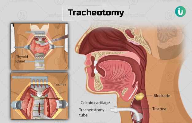
- The surgeon will make a small, horizontal opening in your skin on the front of your neck.
- He/she will separate the muscles in this area and move them to a side until the trachea (windpipe) and the thyroid gland are seen.
- The surgeon will then pull up the thyroid gland or remove it during the surgery.
- He/she will cut your trachea open vertically or horizontally and create a flap with the tracheal cartilage. Alternatively, the surgeon will remove a section of the anterior wall of your trachea to create a small opening.
- Next, the surgeon will introduce a tracheotomy tube through this opening. He/she will verify the correct placement of the tracheotomy tube through the ease of air exchange, visually checking the placement, and oxygen saturation and exhaled carbon dioxide levels.
- The tube will be kept in place with two sutures on each side, followed by a tracheotomy tape.
Percutaneous dilational technique (bedside tracheotomy)
Bedside tracheotomy is a minimally invasive procedure done under general anaesthesia. In this procedure, the tracheotomy tube is inserted without directly visualising the trachea in an individual who has an endotracheal tube inserted.
- You will be asked to lie down with your neck in an extended position.
- The surgeon will make an incision in the front of your neck and move aside the tissue above the trachea.
- He/she will withdraw the endotracheal tube such that its cuff is at the same level as the glottis (the vocal apparatus that contains the vocal cords).
- Next, the surgeon will insert a fiberoptic bronchoscope through the endotracheal tube so that the light from its tip is seen from the incision.
- The surgeon will examine your trachea by touching the skin above the area He/she will determine the position of entry into the trachea through the bronchoscope light that will be seen through your skin.
- Your doctor will insert a Teflon catheter introducer needle in an appropriate position in the trachea taking care not to injure the back wall of the trachea.
- Once that is done, the surgeon will remove the needle, and dilate its path into the trachea with the help of a dilator. A tracheostomy tube will then be inserted over the dilator.
- The surgeon will check the placement of the tube, and tie the tube in place with sutures on your skin and a tracheostomy tape.
Emergency tracheostomy:
This can be done by two methods – open tracheostomy or cricothyroidotomy.
Open tracheostomy:
This is performed as described above to gain urgent access to the airway.
Cricothyroidotomy: For this procedure an opening is made to the cricothyroid membrane (the membrane covering the cricoid cartilage that lies in front of the thyroid), as this membrane is closest to the surface of the skin, and less dissection is required.
- The surgeon will locate your cricothyroid membrane, insert a knife into the membrane and then twisted vertically to open the membrane. He/she will then place an endotracheal tube in the opening.
- When your condition stabilises or if breathing support is needed for more than 2-4 days, then a conventional tracheostomy is performed.
The doctor will remove the tube when it is no longer required and you are able to breathe on your own. The opening does not need to be stitched as it will heal on its own. Once the tube is removed, a bandage will be placed over the opening. In about two weeks, the opening should close.
You will stay in the hospital for three to five days. You can expect the following when you wake up after the surgery:
- You will have a humidity collar in front of the tracheostomy tube to provide moisturised air rich in oxygen.
- You will have feeding tubes through your nose for food until you can eat.
- Antibiotics may be given to reduce the risk of infection.
- A healthcare practitioner will take your chest X-ray to ensure that the tube is in place and there are no complications. The tracheostomy tube will be changed 7 days after surgery.
- Before discharge, you will be shown how to clean the catheter, manage the suction within the tube, clean the skin around the tracheostomy site, and humidify the air you breathe in.
- You may communicate with others by writing, as your speech may be slightly affected. A speech therapist will help you learn to speak.
- You may require training for swallowing, chewing, and breathing with the tube in place. Nutritionists may help in this process.

