Before the surgery, the patient changes into a hospital gown. Thereafter, the patient is taken into the operating room where they lie in the supine position (on their back) on the operating table. To keep track of vitals, the patient is attached to a monitor. An IV cannula is attached that administers medication during the surgery
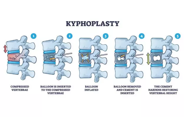
Fluoroscopy is used for X-ray guidance while performing the surgery. A hollow needle (called trocar) is inserted into the skin and passing through the back muscles is guided to the fractured area of the bone. Thereafter, a special balloon (bone tamp) is inserted through the trocar and into the vertebra.
Once in position, the balloon will be gently inflated to create space for a hole or cavity inside the vertebra, thus returning it to its natural height. The balloon is then deflated and removed.
After this, using a specially designed instrument, the cavity that is created is slowly filled with polymethylmethacrylate, which appears like toothpaste and is a cement-like material, also referred to as bone cement. This material hardens typically within 20 minutes.
After the material is injected, the trocar is removed and no stitches are needed. Pressure is put to stop any bleeding and the opening in the skin is covered with a bandage.
The entire procedure usually takes up to one hour. However, if there is more than one vertebra, it may take more time.


